Barrett's esophagus
Board exam relevance: in 2 of 105 exam reports · rank 142- Synonyms
- Barrett oesophagus, Barrett metaplasia, columnar-lined oesophagus
- Specialty
- Internal medicine · Gastroenterology
- Images
- Histology 2
- Last updated
- 10/2026 · Dr. Pascal Bafteh
Contents
Images (2)
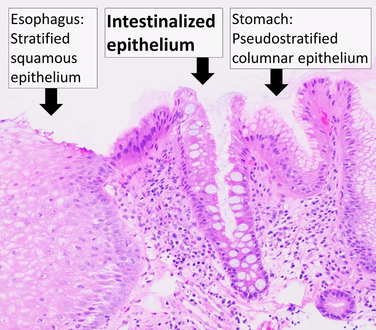 Histology
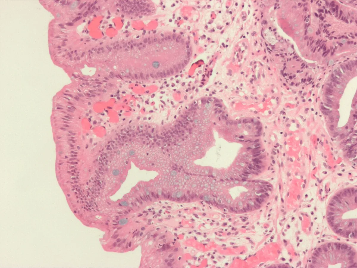 Histology
Definition
Barrett's esophagus is the replacement of the normal squamous epithelium of the distal esophagus by metaplastic columnar epithelium. The new mucosa is glandular and intestine-like, with a brush border and goblet cells.
Barrett's esophagus develops during the healing phase of acute reflux esophagitis when healing takes place in the continued presence of gastric acid. It is regarded as a cancer risk factor: most esophageal adenocarcinomas arise from Barrett's esophagus.
Occurrence & epidemiology
Barrett's esophagus mainly affects people with long-standing gastro-esophageal reflux disease. In Germany, esophageal adenocarcinomas, which frequently arise from Barrett's esophagus, have risen in recent years to about 48 % of all esophageal cancers; in men they now account for the larger share at about 52 %.
Aetiopathogenesis
- Chronic reflux disease and reflux esophagitis: the basis of Barrett's esophagus; metaplasia develops while the mucosa heals under continued acid exposure.
- Obesity: increases the risk of esophageal adenocarcinoma, probably because it promotes reflux.
- Familial clustering: familial Barrett's esophagus is described as a genetic syndrome with an increased cancer risk.
- Cancer development: Barrett's esophagus is a precursor; adenocarcinoma of the distal esophagus can develop via dysplasia.
Clinical features
Barrett's esophagus itself causes no specific symptoms. The symptoms of the underlying reflux disease, such as heartburn and regurgitation, predominate. Dysphagia, loss of weight, signs of bleeding or iron deficiency anemia point to a complication such as a stricture or carcinoma.
Histology
Typical findings are metaplastic, columnar, glandular epithelium of the small-intestinal type with a brush border and goblet cells in place of the stratified squamous epithelium. Histological confirmation of the metaplasia is required for the diagnosis.
Diagnosis
- Endoscopy: red-appearing bands of metaplastic mucosa extending proximally from the esophagogastric junction; described using the Prague classification with circumferential (C) and maximum extent (M). Barrett's esophagus is diagnosed only from a length of 1 cm.
- Biopsy: the only test that consistently detects the columnar mucosal changes of Barrett's esophagus.
- If carcinoma is suspected: biopsy of suspicious lesions, followed by staging (see esophageal cancer).
Keep learning in the app
Further reading (open access)
Cross-references
More topics: Gastroenterology
Note: Learning content for medical education – not a treatment recommendation and no substitute for diagnosis or treatment decisions in individual cases. Treatment and management are deliberately not covered on this page.