Crohn's disease
Board exam relevance: in 7 of 105 exam reports · rank 53- Synonyms
- Crohn disease, regional enteritis, terminal ileitis, IBD, inflammatory bowel disease
- Specialty
- Internal medicine · Gastroenterology
- Images
- CT 1 · MRI 1 · X-ray 1 · Histology 1
- Last updated
- 10/2026 · Dr. Pascal Bafteh
Contents
Images (4)
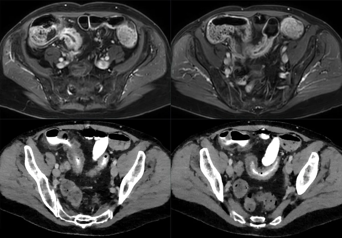 CT
CT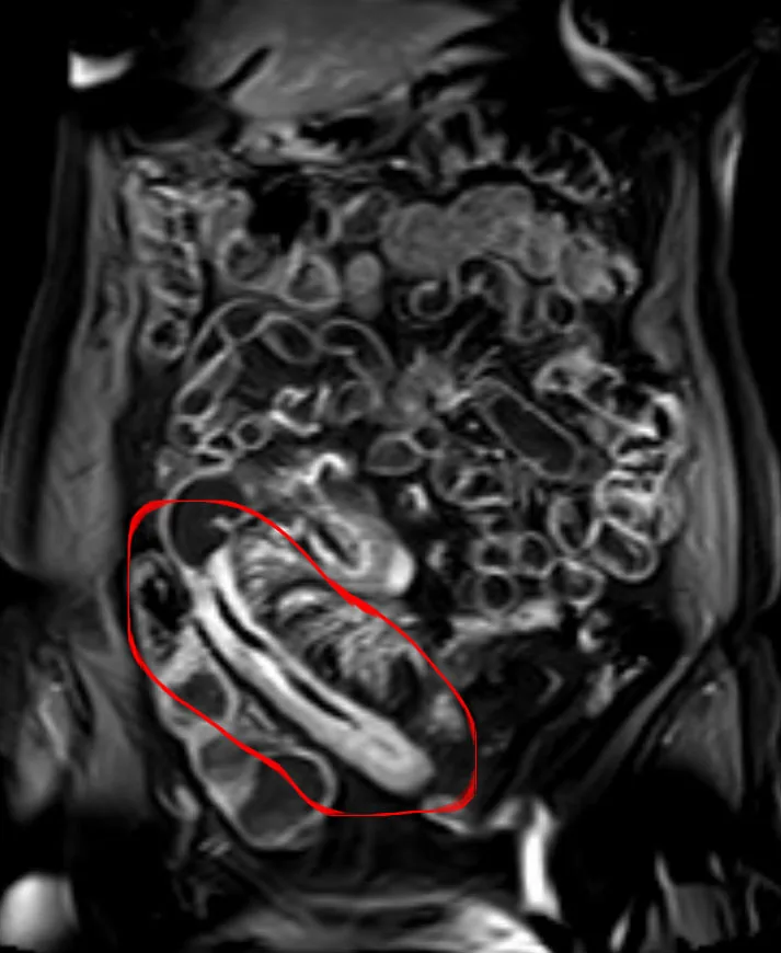 MRI
MRI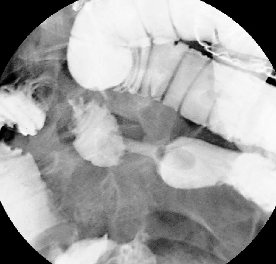 X-ray
X-ray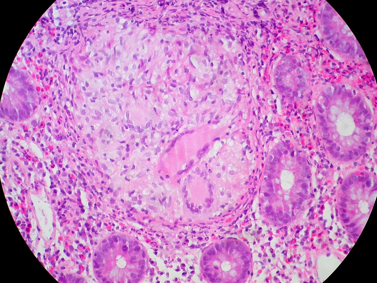 Histology
Definition
Crohn's disease is a chronic transmural inflammatory bowel disease (IBD) that usually affects the distal ileum and colon but may occur in any part of the gastrointestinal tract. The cardinal symptoms are diarrhea and abdominal pain; abscesses, internal and external fistulas and bowel obstruction may arise. Diseased segments are sharply demarcated from adjacent normal bowel (skip areas), hence the name regional enteritis.
Classification
- Pattern of involvement: about one third ileum alone (ileitis), about one third ileum and colon (ileocolitis, predominantly the right colon), about one third colon alone (granulomatous colitis, mostly sparing the rectum). The ileum is inflamed in about 80% of cases; occasionally the small bowel is involved (jejunoileitis), and the stomach, duodenum or esophagus is clinically involved only rarely.
- Behavior patterns: primarily inflammatory, often evolving into stricturing, or primarily penetrating or fistulizing.
Montreal classification (app card)
- A — Age at diagnosis: A1 < 17 yrs / A2 17–40 yrs / A3 > 40 yrs.
- L — Location: L1 ileum, L2 colon, L3 ileocolonic, L4 upper GI.
- B — Behavior: B1 inflammatory, B2 stricturing, B3 penetrating (fistulas/abscesses).
- p — perianal involvement: suffix "p" with perianal fistulas/abscesses.
Occurrence & epidemiology
Inflammatory bowel diseases affect about 0.3% of the population in North America, Oceania and Europe. The peak incidence is between 15 and 30 years, with a smaller second peak between 50 and 70 years. IBD is most common in North America and Northern Europe and two to four times more common in people of Ashkenazi Jewish ancestry. Both sexes are affected about equally. First-degree relatives have a 4- to 20-fold increased risk; familial clustering is much stronger in Crohn's disease than in ulcerative colitis.
Aetiopathogenesis
- Basic mechanism: in people with a multifactorial genetic predisposition (probably involving abnormal epithelial barriers and mucosal immune defenses), the normal intestinal flora triggers an inappropriate cell-mediated immune reaction; cytokines, interleukins and tumor necrosis factor are released.
- Genetics: several gene variants conferring a higher risk of Crohn's disease have been identified.
- Risk factors: smoking contributes to the development and flares of Crohn's disease; also oral contraceptive use, urban living, antibiotic exposure in childhood and vitamin D deficiency. NSAIDs may exacerbate IBD.
- Pathology: the disease begins with crypt inflammation and abscesses that progress to small aphthous ulcers; these develop into deep longitudinal and transverse ulcers with intervening mucosal edema – the typical cobblestone appearance. Transmural inflammation causes lymphedema, wall thickening and fibrosis with strictures; fistulas penetrate into adjacent bowel loops, the bladder, the psoas muscle or as far as the skin.
Clinical features
- Initial presentation: most commonly chronic diarrhea with abdominal pain, fever, anorexia and weight loss; the abdomen is tender, sometimes with a palpable mass or fullness.
- Course: flares at irregular intervals throughout life; recurrences tend to involve the same segment. Severe flares cause intense constant pain, fever and dehydration.
- Rectal bleeding: unusual, except in isolated colonic disease.
- Acute abdomen: may simulate appendicitis or intestinal obstruction.
- Perianal manifestations: in about 20–25%, especially fissures and fistulas, sometimes as the first or most prominent complaint.
- Stenoses: colicky pain, distention, obstipation and vomiting.
- Fistulas: enterovesical fistulas with air bubbles in the urine (pneumaturia), draining cutaneous fistulas; free perforation is unusual.
- Children: extraintestinal manifestations often predominate – arthritis, fever of unknown origin, anemia or growth retardation – while abdominal pain or diarrhea may be absent.
- Complications: malabsorption with vitamin D and B12 deficiency; increased cancer risk in affected small-bowel segments; with colonic involvement, a colorectal cancer risk equal to that of ulcerative colitis of the same extent and duration; rarely toxic colitis.
Extraintestinal manifestations (app card)
- Joints: peripheral arthritis + axial spondyloarthritis.
- Skin: erythema nodosum + pyoderma gangrenosum.
- Eye: uveitis, episcleritis.
- Liver: PSC rare in Crohn's, more common in UC.
Histology
Typical findings are transmural inflammation with a focal distribution and fissuring, as well as non-caseating epithelioid granulomas in lymph nodes, peritoneum, liver and all layers of the bowel wall. Granulomas are pathognomonic when present but are found in only up to about 20% of patients.
Diagnosis
- Suspicion: with inflammatory or obstructive symptoms and with perianal fistulas or abscesses, unexplained arthritis, erythema nodosum, fever, anemia or stunted growth in children; a family history increases the likelihood.
- MR or CT enterography: the preferred procedures in less acute presentations; characteristic strictures or fistulas with separation of bowel loops are virtually diagnostic.
- CT of the abdomen: in an acute abdomen (obstruction, abscesses, fistulas, other causes such as appendicitis).
- Ileocolonoscopy with biopsies and sampling for enteric pathogens in the initial evaluation; upper endoscopy may identify subtle gastroduodenal involvement.
- Intestinal ultrasound: assessment without ionizing rays of the bowel wall, mesentery and adjacent structures, particularly in pregnancy and obesity.
- Capsule endoscopy or CT enteroclysis: when findings are questionable; they show superficial aphthous and linear ulcers.
- Laboratory tests: fecal calprotectin; complete blood count (anemia), electrolytes, nutritional status, stool pathogens including Clostridioides difficile; vitamin D and B12; elevated alkaline phosphatase and gamma-glutamyl transpeptidase with major colonic involvement suggest primary sclerosing cholangitis; leukocytes, ESR and CRP are nonspecific.
Keep learning in the app
Further reading (open access)
Cross-references
More topics: Gastroenterology
Note: Learning content for medical education – not a treatment recommendation and no substitute for diagnosis or treatment decisions in individual cases. Treatment and management are deliberately not covered on this page.