Boerhaave and Mallory-Weiss syndrome
Board exam relevance: in 2 of 105 exam reports · rank 142- Synonyms
- oesophageal rupture, spontaneous oesophageal perforation, Mallory-Weiss tear, esophageal rupture, Boerhaave syndrome, Mallory-Weiss syndrome
- Specialty
- Internal medicine · Gastroenterology
- Images
- CT 2 · X-ray 1
- Last updated
- 10/2026 · Dr. Pascal Bafteh
Contents
Images (3)
 CT
CT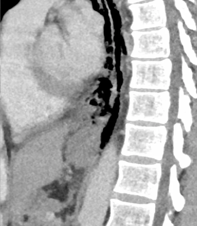 CT
CT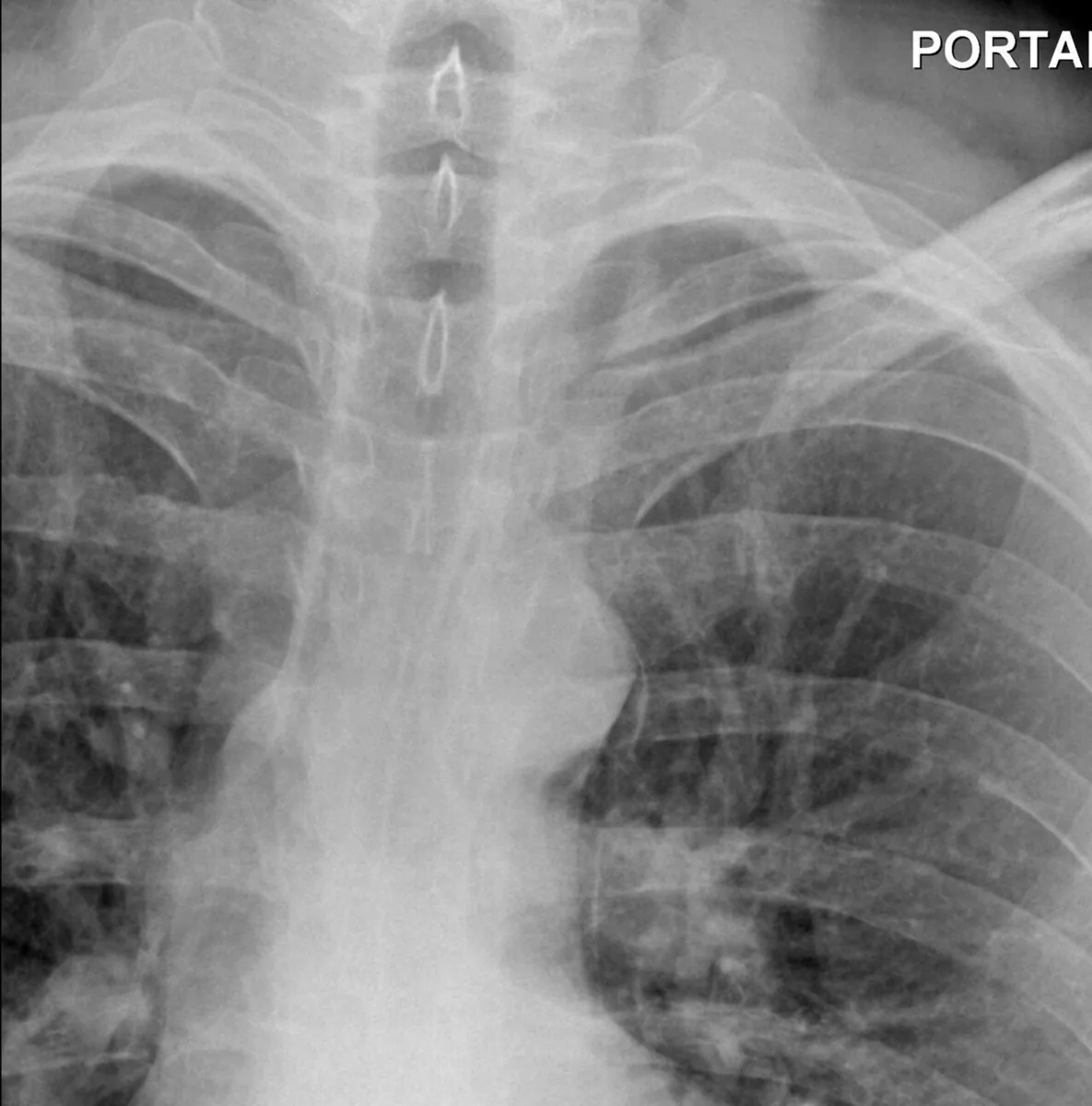 X-ray
X-rayDefinition
Both conditions are linked to a sudden rise in pressure in the esophagus (barotrauma), but they differ in the depth of the injury:
- Mallory–Weiss syndrome: a non-penetrating mucosal laceration of the distal esophagus and proximal stomach caused by vomiting, retching or hiccups.
- Boerhaave syndrome: spontaneous, transmural rupture of the esophagus, typically related to vomiting or retching. Iatrogenic esophageal perforation, e.g. during endoscopy, is to be distinguished.
Occurrence & epidemiology
- Mallory–Weiss syndrome: the cause of about 10 % of upper gastrointestinal bleeds, making it one of the most common causes after variceal and ulcer bleeding. Originally described in people with alcohol use disorder, it can occur with any forceful vomiting.
- Esophageal perforation: in a nationwide study from Iceland, the annual incidence was 3.1 per million population. According to a systematic review, perforations are most often iatrogenic (46.5 %), less often spontaneous (38 %) or due to foreign bodies (6 %); more than 70 % are thoracic.
- Sex and alcohol: both syndromes are more common in men with excessive alcohol consumption.
Aetiopathogenesis
- Mallory–Weiss syndrome: vomiting, retching or hiccups; the tear typically begins just above the squamocolumnar junction and extends proximally.
- Boerhaave syndrome: spontaneous rupture during vomiting, retching or swallowing of a large food bolus; people with uncontrolled eosinophilic esophagitis and acute food impaction are at particular risk.
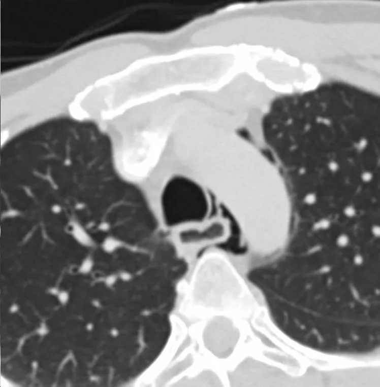
- Consequences of rupture: acid and gastric contents cause fulminant mediastinitis and shock; pneumomediastinum is common, and pleural effusion may also occur.
- Location: most commonly the distal esophagus on the left side.
Clinical features
Mallory–Weiss syndrome:
- typically hematemesis after one or more episodes of non-bloody vomiting
- occasionally lower chest pain
Boerhaave syndrome:
- acute epigastric and retrosternal pain, also abdominal pain, after forceful vomiting
- breathlessness, fever, vomiting, shock
- palpable subcutaneous emphysema in some patients
- Hamman sign: mediastinal crunching synchronous with the heartbeat
Diagnosis
Mallory–Weiss syndrome:
- Clinical picture: typical history of hematemesis after non-bloody vomiting.
- Upper endoscopy: when the history is unclear or bleeding continues; shows a thin, linear tear at the esophagogastric junction.
- Laboratory tests: as part of the work-up of gastrointestinal bleeding.
Boerhaave syndrome:
- CT with water-soluble oral contrast (CT esophagography): the preferred investigation owing to sensitivity, speed and safety.
- Water-soluble contrast esophagogram: an alternative; barium is not used because of possible mediastinal irritation.
- Chest and abdominal X-ray: mediastinal air, pleural effusion or mediastinal widening; Naclerio's V sign (air along the left lower mediastinal border) indicates pneumomediastinum.
- Endoscopy: only in equivocal cases; it may miss a small perforation and may enlarge it.
- Laboratory tests: full blood count (blood loss, sepsis), inflammatory markers, liver and kidney function if sepsis is suspected.
Keep learning in the app
Further reading (open access)
Cross-references
More topics: Gastroenterology
Note: Learning content for medical education – not a treatment recommendation and no substitute for diagnosis or treatment decisions in individual cases. Treatment and management are deliberately not covered on this page.