Eosinophilic esophagitis
Board exam relevance: in 5 of 105 exam reports · rank 69- Synonyms
- EoE, allergic oesophagitis, asthma of the oesophagus, food impaction
- Specialty
- Internal medicine · Gastroenterology
- Images
- Endoscopy 2 · Histology 1
- Last updated
- 10/2026 · Dr. Pascal Bafteh
Contents
Images (3)
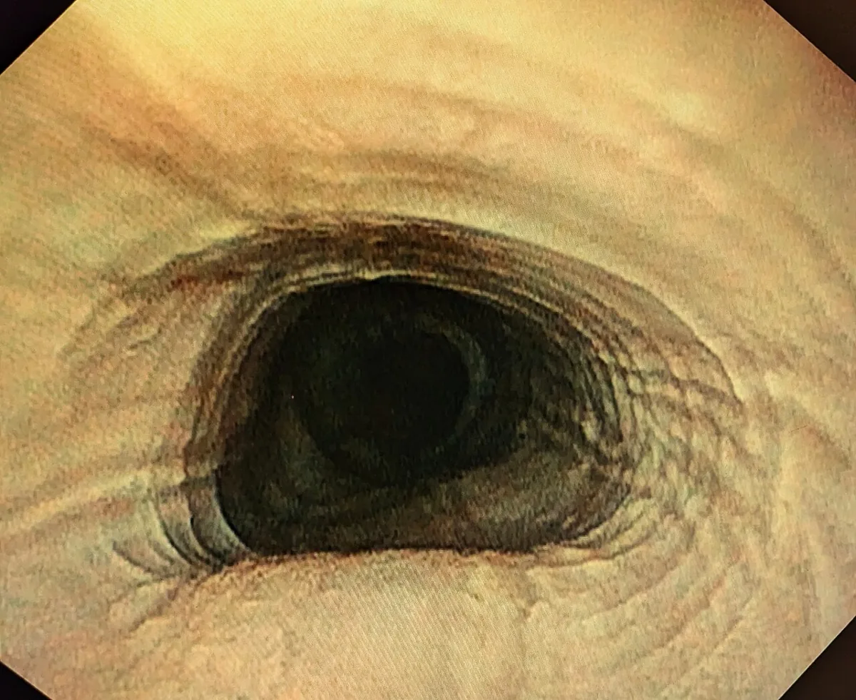 Endoscopy
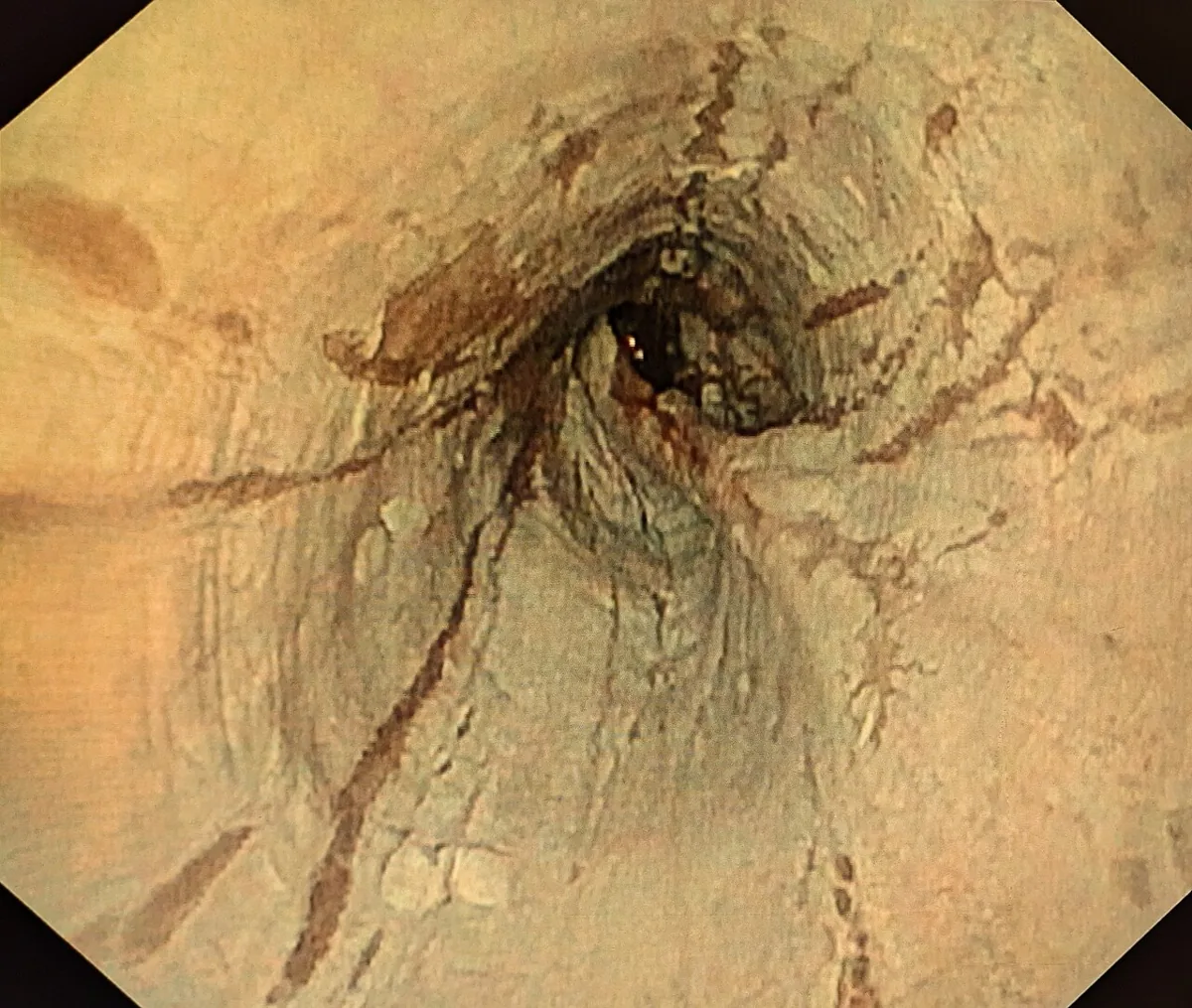 Endoscopy
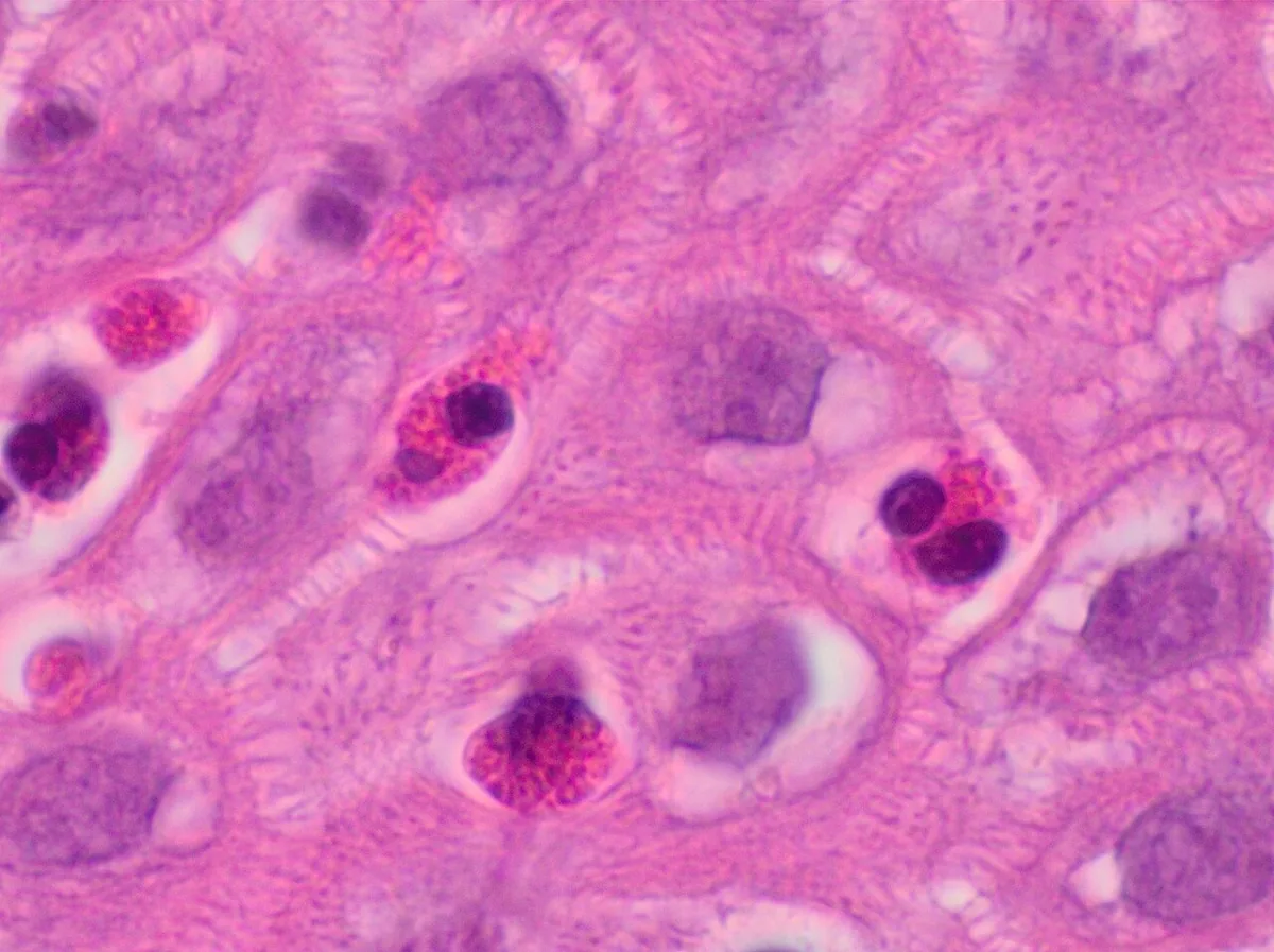 Histology
Definition
Eosinophilic esophagitis (EoE) is a chronic immune-mediated disease of the esophagus with eosinophil-predominant inflammation. It can cause reflux-like symptoms, dysphagia and food impaction. The diagnosis rests on the combination of clinical, endoscopic and histological findings.
Occurrence & epidemiology
EoE was only described as a distinct entity in 1993/1994; incidence and prevalence have increased since then. A meta-analysis found an incidence of 6.6 per 100,000 person-years in children and 7.7 in adults, and a prevalence of 34 per 100,000 children and 42.2 per 100,000 adults. Men are affected about three times as often. More than 65 % of cases occur in childhood, with another peak between 30 and 44 years. The disease is more common in Europe and North America than in Eastern countries.
Aetiopathogenesis
- Immune reaction to food antigens in genetically susceptible individuals; environmental allergens probably also play a role.
- Most common triggers: milk, wheat, soy, eggs, peanuts and tree nuts, and fish and shellfish.
- Type 2 inflammation: Th2 lymphocytes and cytokines such as interleukin-4, -5 and -13; interleukin-13 promotes the production of eotaxin-3, which attracts eosinophils.
- Barrier dysfunction: reduced filaggrin and desmoglein-1 make it easier for allergens to penetrate.
- Genetic risk variants: e.g. in CCL26 (eotaxin-3), TSLP, filaggrin, desmoglein-1, STAT6 and CAPN14.
- Remodelling: persistent inflammation leads to fibrosis and tissue remodelling with narrowing and strictures.
Clinical features
- Infants and young children: vomiting, food refusal, dysphagia, loss of weight, abdominal or chest pain, less often failure to thrive.
- School-age children and adolescents: dysphagia and food impaction, especially with coarse food; also chest and abdominal pain, vomiting, regurgitation.
- Adults: dysphagia for solids and food impaction as the most common symptoms, as well as heartburn.
- Coexisting atopy: frequently asthma, atopic dermatitis or allergic rhinitis.
- Course: chronic; persistent inflammation can lead to narrowing and strictures of the esophagus.
Histology
The histological diagnosis is established with at least 15 eosinophils per high-power field (HPF). Because eosinophilic infiltrates are patchy, biopsies are taken from the upper, middle and lower thirds of the esophagus.
Diagnosis
- Suspicion: typically dysphagia for solids with a history of atopy; also with reflux symptoms that do not improve with acid-suppressing drugs, and in adults with food impaction or non-cardiac chest pain.
- Endoscopy: longitudinal furrows, concentric rings, whitish exudates, strictures, a narrowed lumen, superficial tears on passing the endoscope, loss of vascular markings. The EREFS score summarises rings, furrows, exudates and edema plus additional findings. In 10–25 % the esophagus looks normal at endoscopy – biopsies are therefore always taken.
- Biopsies: 6 to 9 samples from different levels markedly increase sensitivity; samples from the proximal/middle and distal esophagus are processed separately, as reflux disease can also cause eosinophilic infiltrates.
- Barium swallow: may show stacked rings, longitudinal furrows, a narrow calibre or strictures.
- Impedance planimetry: occasionally used to detect subtle strictures.
- Food allergy testing: of little benefit, as EoE is not fully IgE-mediated.
Keep learning in the app
Further reading (open access)
Cross-references
More topics: Gastroenterology
Note: Learning content for medical education – not a treatment recommendation and no substitute for diagnosis or treatment decisions in individual cases. Treatment and management are deliberately not covered on this page.