Ulcerative colitis
Board exam relevance: in 11 of 105 exam reports · rank 24- Synonyms
- UC, colitis, IBD, inflammatory bowel disease, ulcerative proctitis, pancolitis
- Specialty
- Internal medicine · Gastroenterology
- Images
- Endoscopy 1 · X-ray 2 · Histology 1 · Gross specimen 1
- Last updated
- 10/2026 · Dr. Pascal Bafteh
Contents
Images (5)
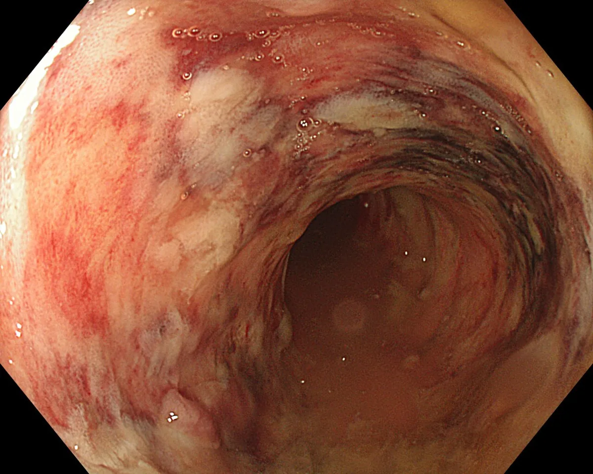 Endoscopy
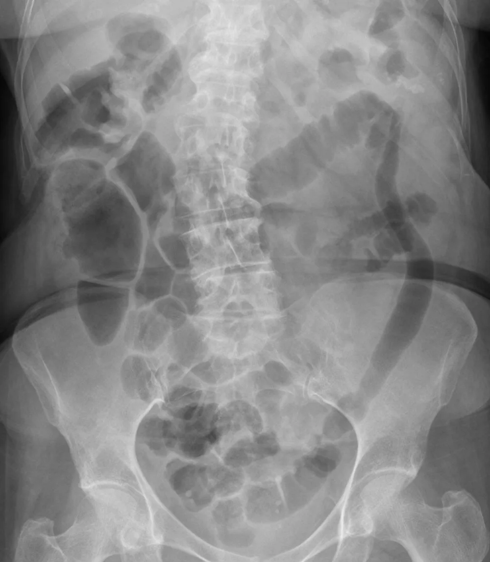 X-ray
X-ray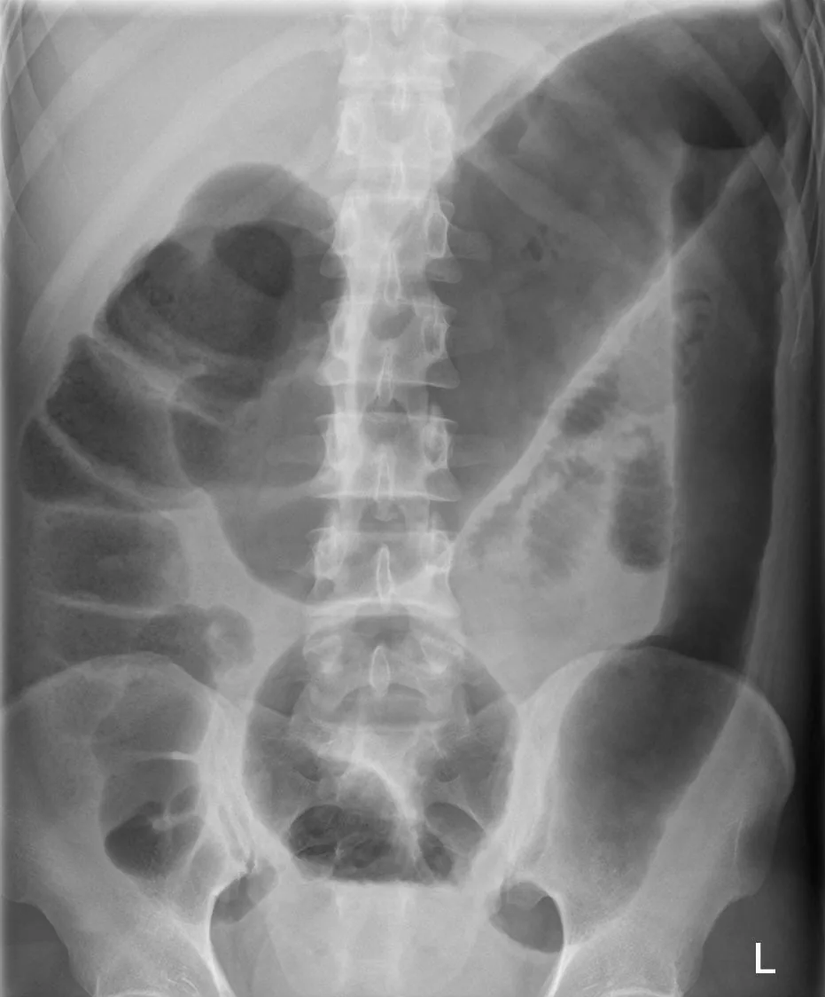 X-ray
X-ray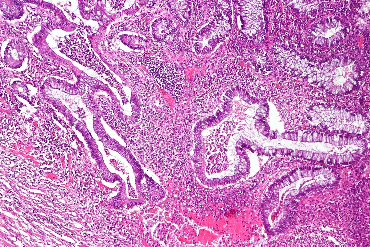 Histology
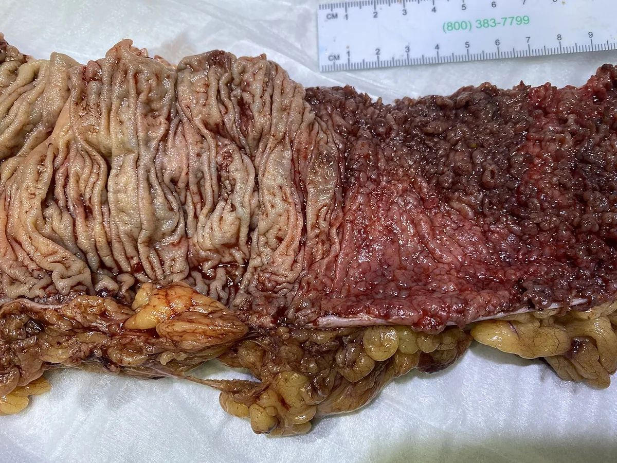 Gross specimen
Definition
Ulcerative colitis (UC) is a chronic inflammatory and ulcerative disease arising in the colonic mucosa, most often characterized by bloody diarrhea. It almost always begins in the rectum and may remain confined to the rectum (ulcerative proctitis) or extend proximally to involve the entire colon. Inflammation affects the mucosa and submucosa with a sharp border between normal and affected tissue; only in severe disease is the muscularis involved.
Together with Crohn's disease it belongs to the inflammatory bowel diseases (IBD). About 5–11% of colitis cases cannot be assigned with certainty to either form and are termed unclassified.
Classification
Distribution pattern (app card)
- UC distribution pattern: continuous from rectum, only mucosa/submucosa.
- Montreal extent E3: pancolitis beyond splenic flexure.
- Typical histology: crypt architecture distortion + crypt abscesses.
Mayo score (app card)
- 4 components (each 0–3 pts, total 0–12): stool frequency + rectal bleeding + endoscopy + physician global assessment.
- Partial Mayo (without endoscopy): 3 components 0–9, usable outpatient.
Acute severe flare (app card)
- Acute severe flare (ASUC) — definition: ≥ 6 bloody stools/day + systemic signs (fever/tachycardia/anemia/ESR↑).
Extent and severity
- Extent (Montreal classification): E1 proctitis (confined to the rectum, distal to the rectosigmoid junction), E2 left-sided colitis (involvement up to the splenic flexure), E3 extensive colitis (beyond the splenic flexure up to pancolitis).
- Severity: the Mayo score uses stool frequency, amount of blood in the stool, a physician global assessment and endoscopic appearance to classify disease as mild, moderate or severe.
- Acute severe ulcerative colitis (Truelove and Witts criteria): at least 6 grossly bloody stools per day with signs of systemic inflammation: fever (mean evening temperature above 37.5 °C or above 37.8 °C on at least 2 of 4 days), pulse above 90/min, anemia with hemoglobin below 75% of normal and ESR above 30 mm/h.
- Toxic (fulminant) colitis: transmural extension of ulceration with localized ileus and peritonitis; megacolon is defined as a transverse colon diameter of more than 6 cm during an exacerbation; the toxic state can also occur without megacolon.
Occurrence & epidemiology
Inflammatory bowel diseases affect about 0.7% of the US population and about 0.3% of the population in North America, Oceania and Europe overall. The peak incidence is between 15 and 30 years; a smaller second peak between 50 and 70 years may include some cases of ischemic colitis. IBD is most common in North America and Northern Europe, less common in Eastern and Southern Europe and rarer still in South America, Asia and Africa. It is two to four times more common in people of Ashkenazi Jewish ancestry. Both sexes are affected about equally.
First-degree relatives have a 4- to 20-fold increased risk (absolute risk about 5–7%); familial clustering is weaker in ulcerative colitis than in Crohn's disease.
Aetiopathogenesis
- Basic mechanism: in people with a multifactorial genetic predisposition – probably involving abnormal epithelial barriers and mucosal immune defenses – the normal intestinal flora triggers an inappropriate cell-mediated immune reaction in the mucosa, with release of cytokines, interleukins and tumor necrosis factor.
- No single cause: specific environmental, dietary or infectious causes have not been identified; however, some cases develop after an intestinal infection (e.g. amebiasis, bacillary dysentery).
- Smoking: current smoking decreases the risk of ulcerative colitis but contributes to Crohn's disease.
- Appendectomy: appendectomy performed for appendicitis appears to lower the risk of ulcerative colitis.
- Other risk factors for IBD: oral contraceptive use, urban living, antibiotic exposure in childhood, vitamin D deficiency; breastfeeding, H. pylori infection and a high folate level appear to be protective.
- Triggers of flares: NSAIDs may exacerbate IBD.
Clinical features
Cardinal symptoms and course
- Typical course: attacks of bloody diarrhea of varied intensity and duration, interspersed with asymptomatic intervals.
- Onset of an attack: usually insidious, with increased urgency to defecate, mild lower abdominal cramps, and blood and mucus in the stools.
- Proctitis or rectosigmoid disease: stool may be normal or hard and dry; rectal discharges of mucus loaded with red and white blood cells accompany or occur between bowel movements; systemic symptoms are absent or mild.
- Extensive ulceration: looser stools, sometimes more than 10 bowel movements per day, severe cramps and distressing rectal tenesmus, also at night; stools watery or mucous, often consisting almost entirely of blood and pus.
- Systemic symptoms: especially with extensive disease, malaise, fever, anemia, anorexia and weight loss.
Complications
- Toxic (fulminant) colitis: sudden violent diarrhea, fever up to 40 °C, abdominal pain, signs of peritonitis (rebound tenderness) and profound toxemia. Within hours to days the colon loses muscular tone and begins to dilate. It usually occurs spontaneously in very severe colitis but can be precipitated by opioid or anticholinergic antidiarrheal drugs.
- Perforation: possible in the course of toxic colitis.
- Colon cancer: risk rises with disease duration and the extent of colon affected, not necessarily with the clinical severity of attacks; sustained microscopic inflammation is considered a risk factor. With coexisting primary sclerosing cholangitis, the risk is increased from the time of diagnosis.
- Pseudopolyps: islands of relatively normal or hyperplastic inflammatory mucosa projecting above ulcerated areas.
- No fistulas or abscesses (unlike Crohn's disease); significant perianal lesions do not occur.
Extraintestinal manifestations
Extraintestinal manifestations are more common in ulcerative colitis and Crohn colitis than in Crohn's disease limited to the small bowel:
- Parallel to intestinal activity: peripheral arthritis (large joints, migratory and transient), episcleritis, aphthous stomatitis, erythema nodosum.
- Independent of intestinal activity: ankylosing spondylitis, sacroiliitis, uveitis, pyoderma gangrenosum and primary sclerosing cholangitis.
- Joint and skin involvement is most common when systemic symptoms are present.
Histology
Inflammation is uniform and diffuse and confined to the mucosa except in severe cases. Endoscopically and pathologically, the mucosa is early on erythematous, finely granular and friable, with loss of the normal vascular pattern and often scattered hemorrhagic areas; large mucosal ulcers with copious purulent exudate characterize severe disease. Typical epithelioid granulomas do not occur.
Diagnosis
History, laboratory and stool tests
- Suspicion: typical symptoms, especially with extraintestinal manifestations or previous similar attacks.
- Stool tests: Clostridioides difficile toxin in all suspected cases, stool cultures for enteric pathogens, fresh stool for Entamoeba histolytica; in immunosuppressed patients also opportunistic infections (e.g. cytomegalovirus).
- Laboratory tests: anemia, hypoalbuminemia, electrolyte abnormalities, systemic inflammation (ESR, CRP), thrombocytosis and leukocytosis; liver tests, with elevated alkaline phosphatase and gamma-glutamyl transpeptidase suggesting primary sclerosing cholangitis.
- Fecal calprotectin and lactoferrin: help differentiate IBD from functional diarrhea; calprotectin is used mainly as a marker of disease activity.
- Serology: pANCA and ASCA do not reliably differentiate ulcerative colitis from Crohn's disease and are not part of routine diagnosis.
Endoscopy
- Colonoscopy with examination of the terminal ileum: when safe, by an experienced operator; allows visual confirmation of colitis, sampling of stool or mucus and biopsies.
- Flexible sigmoidoscopy with biopsies: instead of colonoscopy in severe disease with an increased risk of perforation.
- Typical findings: symmetric, uninterrupted inflammation starting in the rectum with a uniform, diffuse appearance; edema, loss of vascularity, friability, erosions and ulcers.
- Features pointing to Crohn's disease: severe perianal disease, rectal sparing, absence of bleeding and asymmetric or segmental involvement.
- Histology: acute, self-limited infectious colitis can usually be distinguished from chronic ulcerative colitis or Crohn colitis.
Imaging
- Abdominal X-ray: not diagnostic and of low yield, therefore not a routine test; possible findings are mucosal edema, loss of haustration and absence of formed stool in the diseased bowel.
- Acute severe attack: flat and upright abdominal radiographs may show megacolon or gas accumulated over a long, paralyzed segment of colon; careful sigmoidoscopy to assess severity.
- Long-standing disease: shortened, rigid colon with atrophic or pseudopolypoid mucosa.
Keep learning in the app
Further reading (open access)
- MSD Manual Professional: Ulcerative Colitis
- MSD Manual Professional: Overview of Inflammatory Bowel Disease
- StatPearls: Ulcerative Colitis
- DGVS: Aktualisierte S3-Leitlinie Colitis ulcerosa (Version 7.0, AWMF 021-009)
- PMC: C-reactive Protein and Partial Mayo Score in Ulcerative Colitis (Inflamm Bowel Dis 2023)
- PMC: Early-Phase Partial Mayo Score and Endoscopic Improvement (Inflamm Intest Dis 2023)
Cross-references
More topics: Gastroenterology
Note: Learning content for medical education – not a treatment recommendation and no substitute for diagnosis or treatment decisions in individual cases. Treatment and management are deliberately not covered on this page.