Diverticular disease and diverticulitis
Board exam relevance: in 4 of 105 exam reports · rank 90- Synonyms
- diverticulosis, diverticular disease, colonic diverticula, left-sided appendicitis, sigmoid diverticulitis, diverticular bleeding
- Specialty
- Internal medicine · Gastroenterology
- Images
- CT 1 · Endoscopy 1 · Ultrasound 1 · Gross specimen 1
- Last updated
- 10/2026 · Dr. Pascal Bafteh
Contents
Images (4)
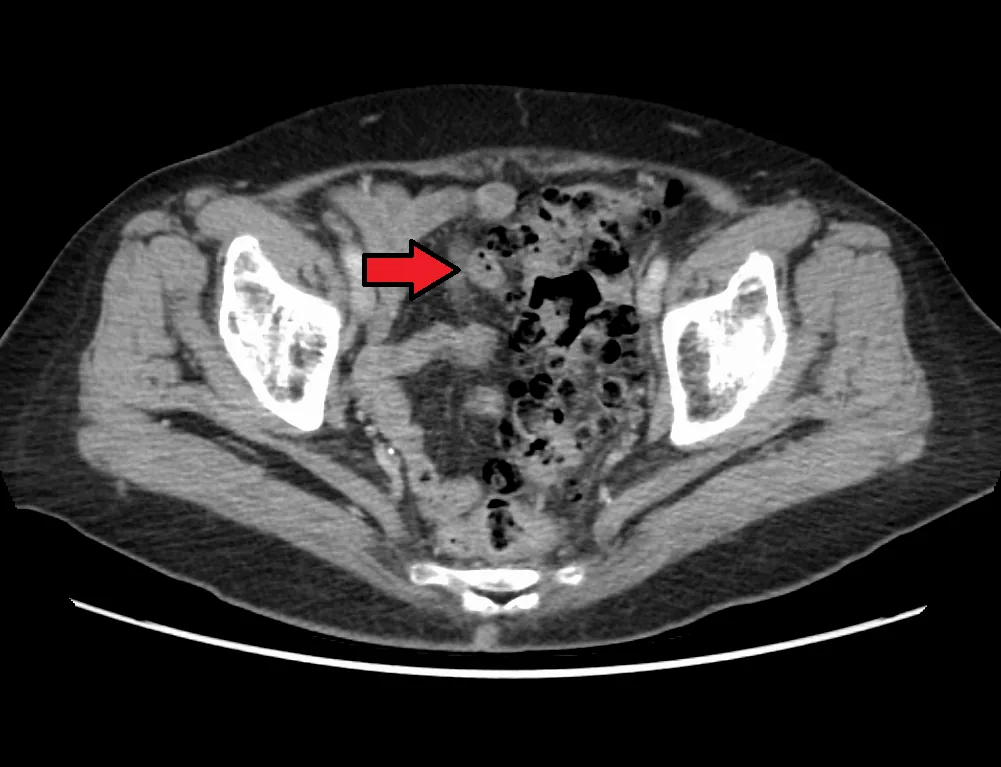 CT
CT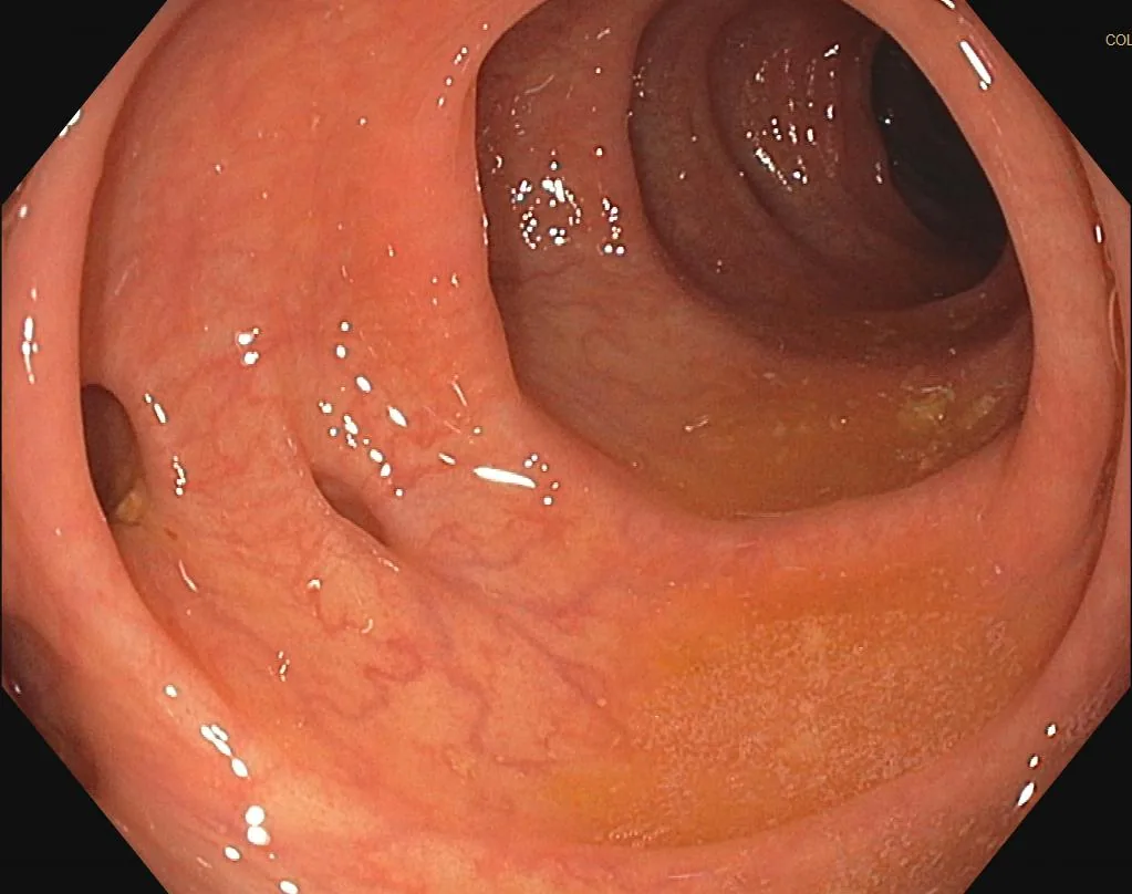 Endoscopy
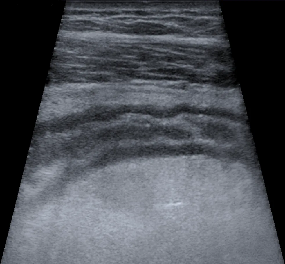 Ultrasound
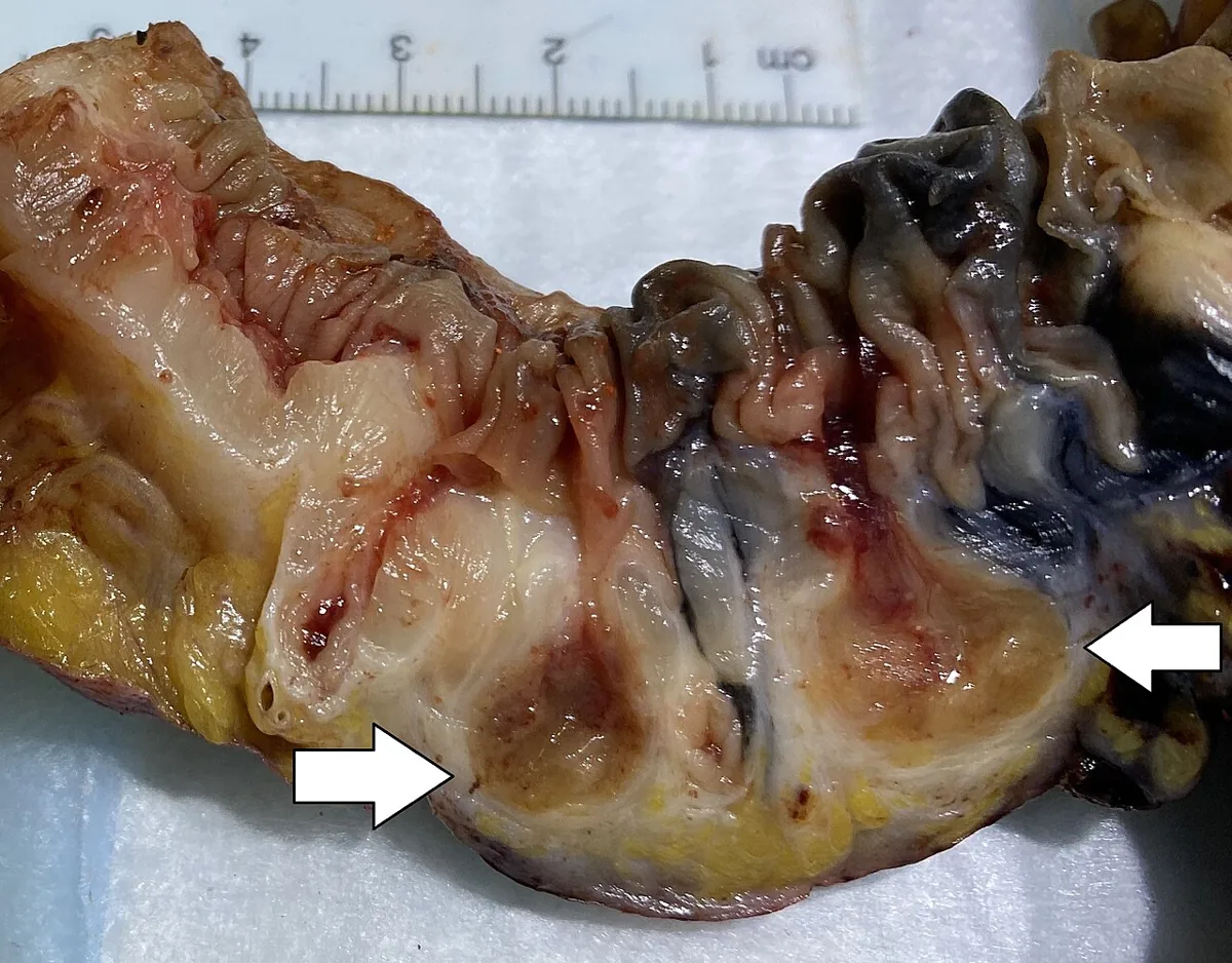 Gross specimen
Definition
Diverticula are saclike mucosal pouches protruding from a hollow organ. True diverticula contain all layers of the wall. Colonic diverticula, by contrast, are pseudodiverticula: mucosa and submucosa protrude through the muscular layer of the bowel wall.
- Diverticulosis: presence of one or more diverticula
- Diverticular disease: symptomatic diverticulosis
- Diverticulitis: inflammation of one or more diverticula, with or without infection
Classification
Hansen-Stock classification
Former German classification; diverticulitis with accompanying phlegmon is already considered complicated here.
- Stage 0: asymptomatic diverticulosis
- Stage I: acute uncomplicated diverticulitis
- Stage IIa: phlegmonous diverticulitis
- Stage IIb: abscess-forming diverticulitis, covered perforation
- Stage IIc: free perforation
- Stage III: chronic recurrent diverticulitis
CDD classification (German S3 guideline)
- Type 0: asymptomatic diverticulosis (incidental finding)
- Type 1: acute uncomplicated diverticular disease/diverticulitis; 1a without, 1b with phlegmonous surrounding reaction
- Type 2: acute complicated diverticulitis; 2a microabscess (covered perforation, abscess up to 3 cm, minimal pericolic air), 2b macroabscess (over 3 cm), 2c free perforation with 2c1 purulent and 2c2 fecal peritonitis
- Type 3: chronic diverticular disease; 3a persistent or recurrent symptoms attributed to diverticulosis (SUDD), 3b recurrent diverticulitis without, 3c with complications (stricture, fistula, conglomerate)
- Type 4: diverticular bleeding
Internationally, acute diverticulitis is classified as uncomplicated (the most common form) or complicated with abscess, fistula, obstruction or free perforation. If it does not heal completely, chronic diverticulitis develops: uncomplicated with wall thickening or chronic mucosal inflammation without stricture, complicated with stenosis and fistulas.
Occurrence & epidemiology
The frequency of diverticulosis increases with age and varies widely by region: half of people aged 60 and older and about 70% of those aged 80 and older have diverticula. In Western countries most diverticula are in the distal (left) colon, especially the sigmoid, and in Asia mostly in the proximal (right) colon. Diverticula are usually 5–10 mm in size.
About 75% of people with diverticulosis remain asymptomatic or have only intermittent constipation; about 25% develop pain or bleeding due to complications. Diverticulitis develops in about 1–5% of people with diverticulosis; about 12% of cases are complicated. Over the age of 50, women are affected more often. Diverticular bleeding occurs in 10–15% of people with diverticulosis and, at 30–65%, is the most common cause of acute lower gastrointestinal bleeding in adults.
Aetiopathogenesis
The pathogenesis is multifactorial and not fully understood. Spasms of the muscular layer probably raise intraluminal pressure so that the mucosa is pushed outward at the weakest points – next to the entry sites of blood vessels. Hereditary factors and alterations of wall structure and motility play a role.
Associated with symptomatic diverticular disease:
- low-fiber eating habits, high intake of red meat
- physical inactivity, obesity, smoking
- intake of certain drugs, including NSAIDs and opioids
Consumption of nuts, seeds, corn or popcorn is not related to the risk. Complications are more common in smokers, obesity, NSAID use and immunodeficiency (e.g. advanced HIV infection). Right-sided diverticula in Western patients have wider necks and thinner domes; the supplying vessels (vasa recta) are stretched more there, which explains the higher bleeding risk.
Clinical features
Acute diverticulitis: left lower quadrant pain and tenderness, often with a palpable sigmoid colon, occasionally suprapubic; in right-sided diverticulitis, right-sided pain. Accompanying nausea, vomiting, fever and sometimes urinary symptoms due to bladder irritation. Peritoneal signs (rebound, guarding) mainly suggest abscess or free perforation. Recurrences present similarly to the first episode and are not necessarily more severe.
Complications: abscess, fistula, obstruction and perforation with peritonitis and sepsis. Fistulas manifest as pneumaturia, fecaluria, feculent vaginal discharge or infection of the abdominal wall, perineum or thigh; colovesical fistulas are more common in women after hysterectomy.
Diverticular bleeding: painless passage of fresh or maroon blood, usually moderate to severe because an arteriole bleeds; usually without concomitant diverticulitis; in most cases it stops spontaneously, and rebleeding is common.
Diagnosis
- CT of the abdomen and pelvis (preferably with contrast): sensitivity 95–99%, specificity 95–100%; bowel wall thickening and inflammatory stranding of the surrounding fat; in about 10% diverticulitis cannot be distinguished from colon cancer on CT
- Ultrasound: an alternative, especially in the outpatient setting; sensitivity 77–98% and specificity 80–99%, highly operator-dependent; less sensitive (55%) for distinguishing complicated from uncomplicated disease
- MRI: an alternative in pregnant and young patients
- Laboratory tests: CRP to estimate the course, complete blood count if sepsis is suspected
- Fecal calprotectin is usually elevated in symptomatic uncomplicated diverticular disease
- Colonoscopy only after the inflammation has resolved, among other things to exclude cancer; diverticula are also found incidentally at colonoscopy, capsule endoscopy, CT or MRI
Keep learning in the app
Further reading (open access)
Cross-references
Note: Learning content for medical education – not a treatment recommendation and no substitute for diagnosis or treatment decisions in individual cases. Treatment and management are deliberately not covered on this page.