Peptic ulcer disease
Board exam relevance: in 8 of 105 exam reports · rank 39- Synonyms
- stomach ulcer, gastric ulcer, duodenal ulcer, peptic ulcer, PUD
- Specialty
- Internal medicine · Gastroenterology
- Images
- Endoscopy 2 · X-ray 1
- Last updated
- 10/2026 · Dr. Pascal Bafteh
Contents
Images (3)
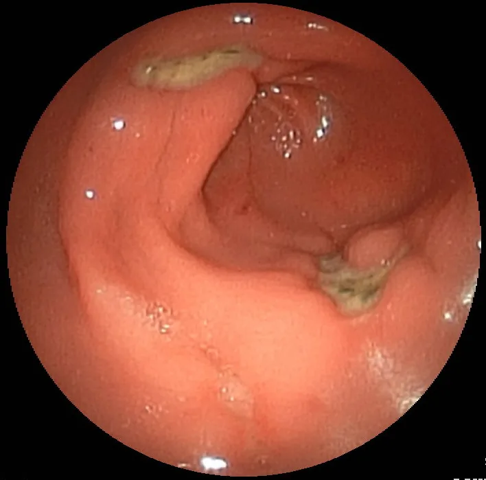 Endoscopy
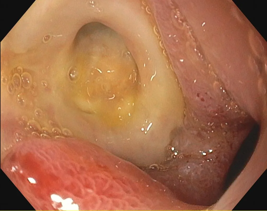 Endoscopy
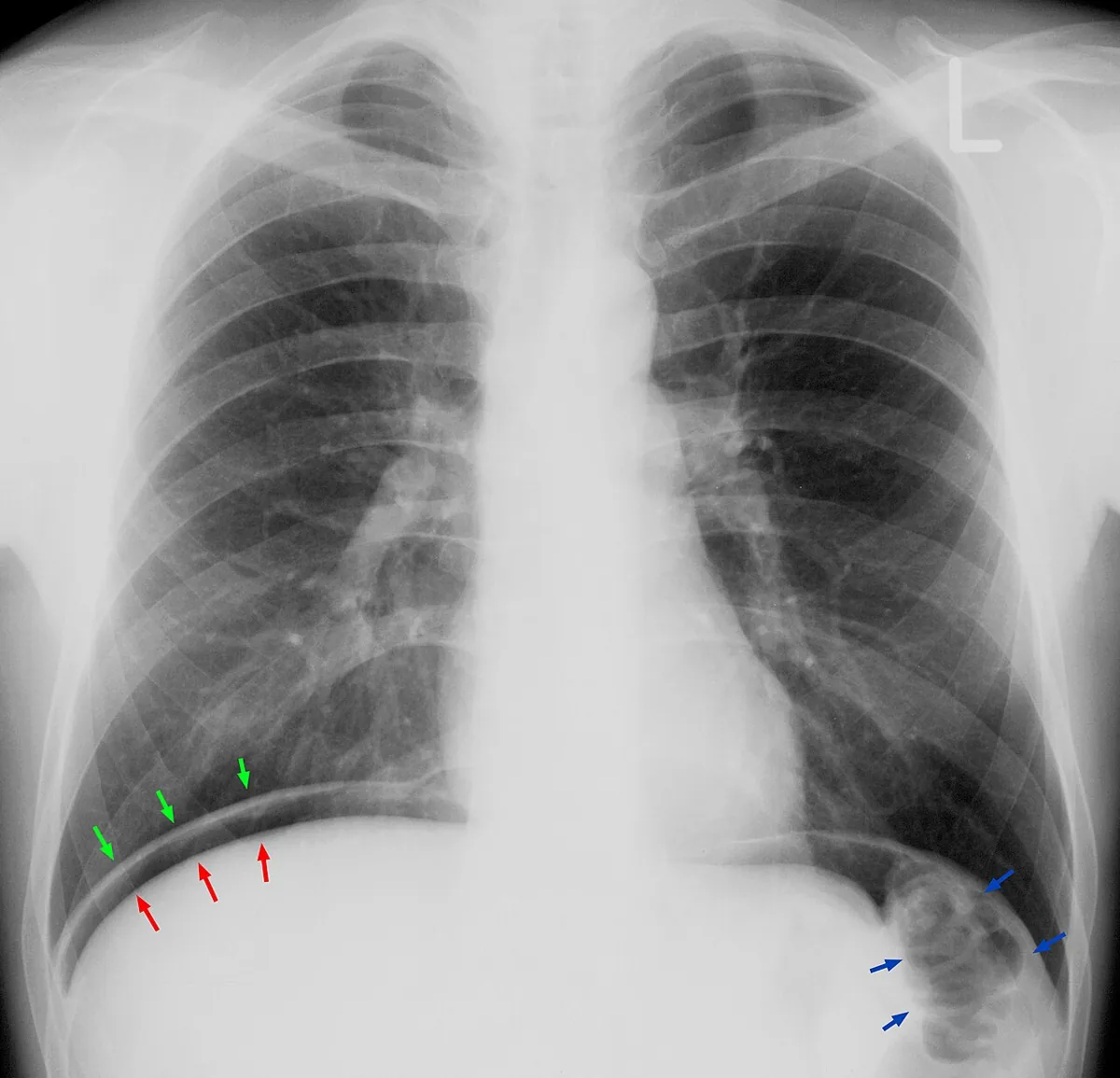 X-ray
X-rayDefinition
A peptic ulcer is a mucosal defect of the gastrointestinal tract, typically in the stomach (gastric ulcer) or the first few centimetres of the duodenum (duodenal ulcer), that penetrates through the muscularis mucosae. Ulcers range from a few millimetres to several centimetres in size. Erosions are more superficial and do not involve the muscularis mucosae.
Occurrence & epidemiology
Ulcers occur at any age, including infancy and childhood, but are most common in middle-aged adults.
Aetiopathogenesis
Nearly all ulcers are caused by Helicobacter pylori infection or by NSAIDs; both disrupt normal mucosal defence and repair and make the mucosa more susceptible to acid.
- H. pylori: present in over 50 % of duodenal ulcers and 30–50 % of gastric ulcers.
- NSAIDs: now account for more than 50 % of peptic ulcers.
- Smoking: a risk factor for ulcers and their complications; risk rises with the number of cigarettes smoked per day.
- Alcohol: increases acid secretion; a link between moderate amounts and ulcer development has not been established.
- Gastrinoma (Zollinger–Ellison syndrome): a rare cause with gastrin hypersecretion.
Clinical features
- Pain: the most common symptom, usually epigastric, burning or gnawing, sometimes like hunger; often relieved by food. The course is usually chronic and recurrent. Only about half of patients present with the characteristic pattern.
- Duodenal ulcer: no pain on waking, pain from mid-morning, relief after eating and recurrence 2–3 hours after a meal; pain that wakes the patient at night is highly suggestive of duodenal ulcer.
- Gastric ulcer: inconsistent pattern, eating may even worsen the pain; pyloric channel ulcers often cause symptoms of obstruction (bloating, nausea, vomiting).
- Older people: often few or no symptoms.
- Neonates and young children: perforation or hemorrhage may be the first sign.
- Hemorrhage (most common complication): hematemesis (fresh blood or coffee grounds), hematochezia or melaena, weakness, orthostasis, syncope, thirst and sweating.
- Penetration (confined perforation): into the lesser sac or adjacent organs such as the pancreas or liver; intense, persistent, position-dependent pain, radiating to the back when a posterior duodenal ulcer penetrates the pancreas.
- Free perforation: usually in the anterior wall of the duodenum; sudden, intense, continuous epigastric pain that spreads rapidly across the abdomen, sometimes referred to the shoulder; board-like rigidity, rebound tenderness, reduced bowel sounds, shock.
Diagnosis
- Endoscopy: confirms the diagnosis; biopsy or brush cytology of gastric lesions to distinguish simple ulceration from ulcerating gastric cancer – especially in people over 60, with loss of weight, or with severe or persistent symptoms. Duodenal ulcers are generally not biopsied because malignancy is extremely rare.
- H. pylori testing: whenever an ulcer is detected.
- Serum gastrin: with multiple, atypically located (e.g. postbulbar) or refractory ulcers, or with prominent diarrhea or loss of weight (suspected gastrinoma).
- Penetration: usually confirmed by CT or MRI.
- Free perforation: free air under the diaphragm or in the peritoneal cavity on X-ray or CT; upright views are preferred, the lateral chest view is the most sensitive, and in severely ill patients a lateral decubitus view is used. Absence of free air does not exclude perforation.
Keep learning in the app
Further reading (open access)
Cross-references
More topics: Gastroenterology
Note: Learning content for medical education – not a treatment recommendation and no substitute for diagnosis or treatment decisions in individual cases. Treatment and management are deliberately not covered on this page.