Choledocholithiasis and acute cholangitis
- Synonyms
- bile duct infection, bile duct stones, common bile duct stones, ascending cholangitis, Charcot triad, obstructive jaundice
- Specialty
- Internal medicine · Gastroenterology
- Images
- Clinical 1 · Ultrasound 1 · MRI 1
- Last updated
- 10/2026 · Dr. Pascal Bafteh
Contents
Images (3)
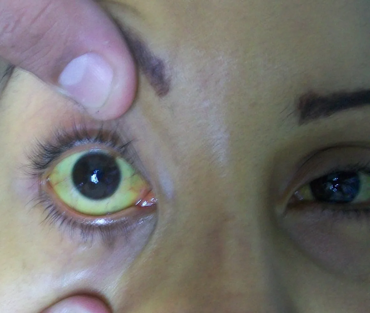
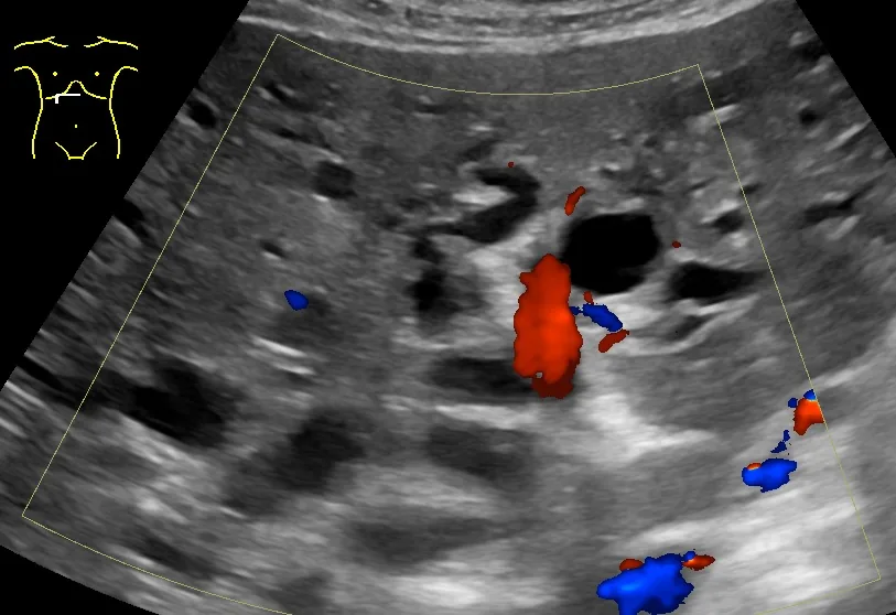 Ultrasound
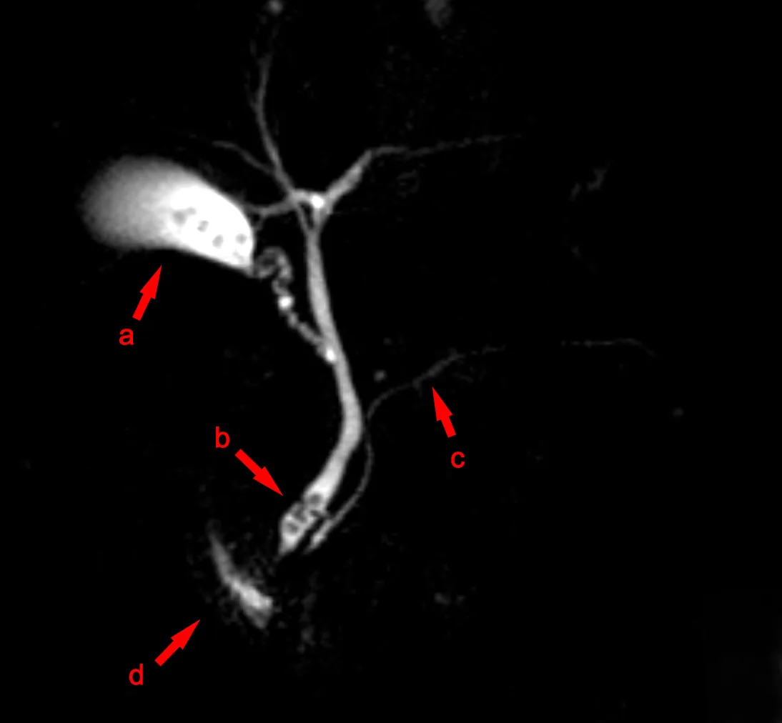 MRI
MRIDefinition
Choledocholithiasis is the presence of stones in the bile ducts; they can form in the gallbladder or in the ducts themselves. Duct stones cause biliary colic, biliary obstruction, gallstone pancreatitis or acute cholangitis, i.e. bacterial infection and inflammation of the bile ducts. Cholangitis in turn can lead to strictures, stasis and further stone formation.
Classification
- Primary stones: usually brown pigment stones that form in the bile ducts
- Secondary stones: usually cholesterol stones that form in the gallbladder and migrate into the ducts
Severity grades according to the Tokyo Guidelines 2018
- Grade III (severe): circulatory failure (shock), impaired consciousness, or failure of the lungs, kidneys, liver or coagulation
- Grade II (moderate): at least two of: white blood cell count above 12 G/L or below 4 G/L, fever of 39 °C or higher, age 75 years or older, total bilirubin of 5 mg/dL (103 µmol/L) or higher, albumin below 25 g/L
- Grade I (mild): no criteria for grade II or III
Occurrence & epidemiology
In industrialized countries, more than 85% of common duct stones are secondary; affected patients also have stones in the gallbladder. Up to 10% of patients with symptomatic gallstones also have common bile duct stones. Duct stones obstructing the ampulla of Vater cause gallstone pancreatitis, the most common cause of acute pancreatitis. About 85% of cases of acute cholangitis result from common bile duct stones.
Aetiopathogenesis
Duct stones may pass into the duodenum asymptomatically. Partial obstruction of the ducts causes biliary colic; more complete obstruction leads to duct dilation, jaundice and eventually ascending cholangitis: the obstruction allows bacteria to ascend from the duodenum. With stasis (e.g. due to a postoperative stricture) and infection, brown pigment stones can also form within the ducts.
Pathogens: mostly gram-negative bacteria (e.g. Escherichia coli, Klebsiella, Enterobacter), less often gram-positive bacteria (e.g. Enterococcus) and mixed anaerobes (e.g. Bacteroides, Clostridia).
Causes of bile duct obstruction:
- stones (common)
- postoperative bile duct injury (common)
- tumors
- scarring due to chronic pancreatitis
- external pressure from cysts, choledochocele or pancreatic pseudocyst (rare)
- strictures in primary sclerosing cholangitis
- late-stage HIV-related cholangiopathy
- parasites (Clonorchis sinensis, Opisthorchis viverrini; rarely Ascaris)
Recurrent pyogenic cholangitis (hepatolithiasis) with intrahepatic brown pigment stones occurs in people of Asian descent; undernutrition and parasitic infestation increase susceptibility, and repeated cycles of obstruction, infection and inflammation lead to strictures and biliary cirrhosis.
Clinical features
Charcot triad and Reynolds pentad
- Charcot triad: right upper quadrant abdominal pain, jaundice, fever or chills; present in only 16 to at most 72% of cases in studies
- Reynolds pentad: Charcot triad plus confusion and hypotension
Choledocholithiasis: biliary colic and jaundice; some patients, usually older ones, present with biliary obstruction without previous symptoms.
Acute cholangitis: abdominal pain, jaundice and fever or chills (Charcot triad); the triad is specific (90%) but not very sensitive. The abdomen is tender, and the liver is often enlarged and tender (possibly containing abscesses). When confusion and hypotension are added, this is called the Reynolds pentad.
Diagnosis
- Suspicion: duct stones with jaundice and biliary colic; fever and leukocytosis additionally suggest cholangitis
- Laboratory tests: elevated bilirubin and especially alkaline phosphatase, ALT and γGT are consistent with extrahepatic obstruction; in cholangitis leukocytosis, aminotransferases sometimes up to 1000 U/l (acute hepatic necrosis, often due to microabscesses)
- Blood cultures and complete blood count are essential if cholangitis is suspected
- Ultrasound: stones in the gallbladder, less often in the common bile duct (less accurate); dilated common bile duct (over 6–7 mm with an intact gallbladder, cut-off depending on the source; over 10 mm without a gallbladder); if the ducts are not dilated early in the course, the stones have probably passed
- MRCP: highly accurate for retained duct stones
- Endoscopic ultrasound or endoscopic ductography if MRCP is equivocal
- CT: less accurate than ultrasound, but detects liver abscesses and duct dilation
Diagnostic criteria and imaging
- Tokyo Guidelines 2018 diagnostic criteria: (A) signs of systemic inflammation (fever; leukocytosis or leukopenia, elevated CRP), (B) obstructive jaundice or elevated cholestasis parameters, (C) on imaging a dilated bile duct, duct stone or other abnormal duct finding (e.g., stricture); definite with A + B + C, suspected with A + B or A + C
- In studies, the Tokyo criteria are clearly superior to the Charcot triad diagnostically
- Transabdominal ultrasound is the first imaging modality
- MRCP and endoscopic ultrasound achieve comparably high sensitivity and specificity for duct stones; MRCP is limited for stones smaller than 5 mm
Keep learning in the app
Further reading (open access)
Cross-references
More topics: Gastroenterology
Note: Learning content for medical education – not a treatment recommendation and no substitute for diagnosis or treatment decisions in individual cases. Treatment and management are deliberately not covered on this page.