Cholangiocarcinoma (bile duct cancer)
Board exam relevance: in 1 of 105 exam reports · rank 181- Synonyms
- bile duct cancer, CCA, Klatskin tumor, intrahepatic cholangiocarcinoma, perihilar cholangiocarcinoma, extrahepatic cholangiocarcinoma
- Specialty
- Internal medicine · Gastroenterology
- Images
- CT 1 · Histology 1 · Gross specimen 1
- Last updated
- 10/2026 · Dr. Pascal Bafteh
Contents
Images (3)
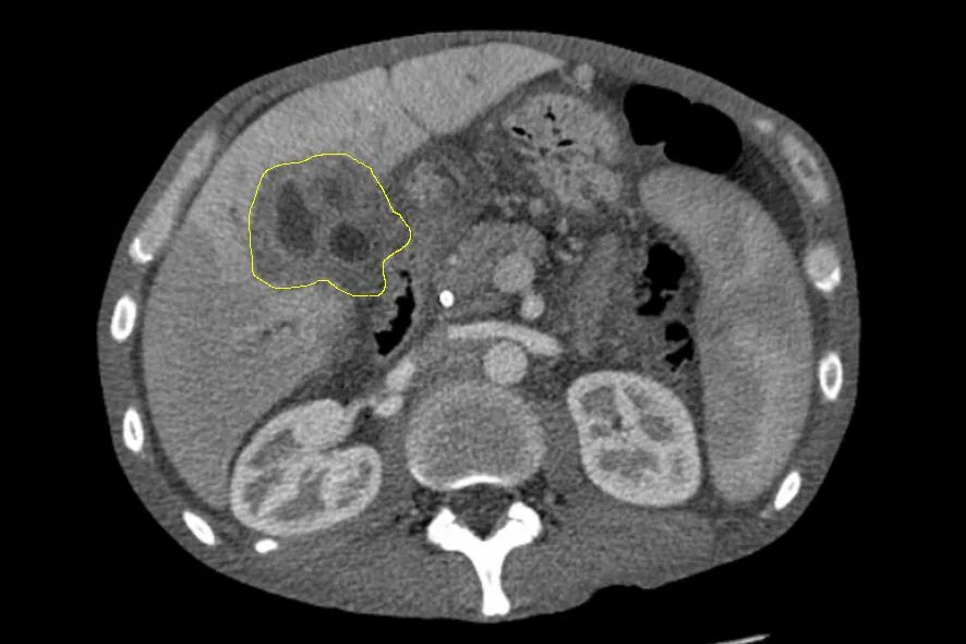 CT
CT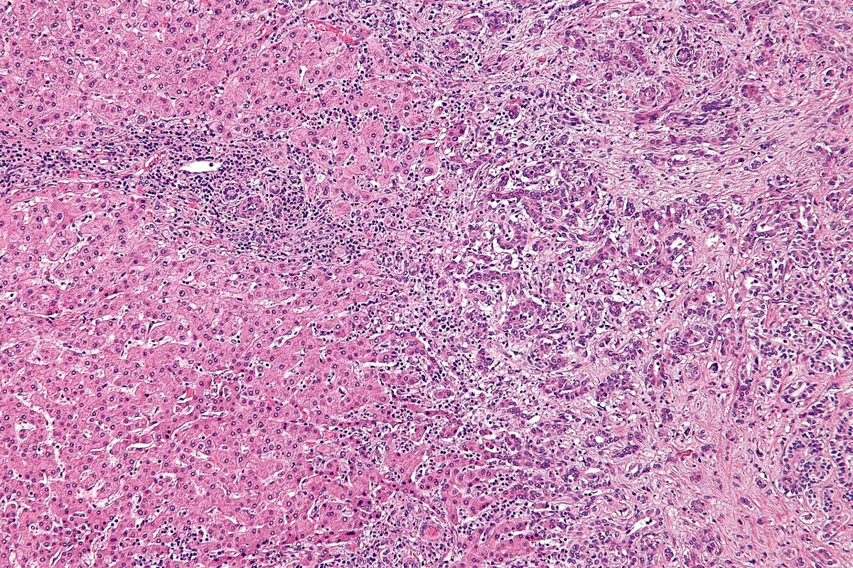 Histology
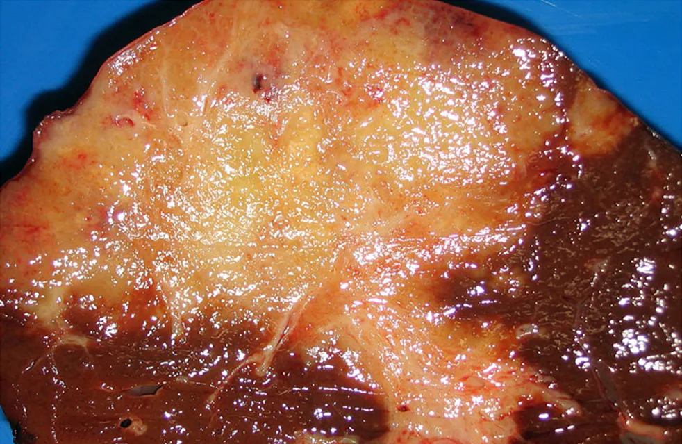 Gross specimen
Definition
Cholangiocarcinoma (CCA, bile duct cancer) comprises a heterogeneous group of malignancies arising in the biliary tree. Histologically, they are adenocarcinomas or mucinous carcinomas with tubular and/or papillary structures and a variable fibrous stroma. CCA is the second most common primary liver malignancy after hepatocellular carcinoma; it accounts for about 15% of primary liver tumors and 3% of gastrointestinal cancers.
Classification
By location:
- intrahepatic (iCCA): above the second-order bile ducts, in the liver periphery
- perihilar (pCCA): in the right and/or left hepatic duct or at their junction (Klatskin tumor)
- distal (dCCA): in the common bile duct; the boundary between perihilar and distal is the insertion of the cystic duct
Perihilar and distal tumors are collectively called extrahepatic CCA; they are considerably more common than intrahepatic tumors, with perihilar tumors forming the largest group.
By growth pattern: mass-forming (mass in the liver parenchyma), periductal-infiltrating (spreading within and along the duct wall) and intraductal-growing (polypoid or papillary into the duct lumen).
Staging: according to the AJCC TNM system.
Occurrence & epidemiology
Cholangiocarcinomas are rare (worldwide incidence about 0.3–6 per 100,000 population per year) but considerably more common in regions with liver flukes such as South Korea, China and Thailand; incidence and death rates have increased worldwide over recent decades. In Germany, about 4,670 malignant tumors of the gallbladder and extrahepatic bile ducts were diagnosed in 2023, about 72% of them in the bile ducts; the risk increases continuously with age.
Aetiopathogenesis
The exact triggers are not clearly identified; the main risk factor is age. Established or possible risk factors:
- primary sclerosing cholangitis and other chronic inflammatory bile duct diseases
- liver flukes (especially in Asia)
- choledochal cysts, congenital biliary anomalies (Caroli syndrome), intrahepatic bile duct stones
- liver cirrhosis, viral hepatitis B and C, alcohol-related liver disease
- severe obesity, diabetes mellitus, smoking
- inflammatory bowel disease
At the molecular level, numerous alterations are found, including in TP53, KRAS, BAP1 and ARID1A, as well as IDH1/IDH2 mutations and FGFR2 fusions in intrahepatic CCA.
Clinical features
Cholangiocarcinomas are usually asymptomatic in early stages and are therefore often diagnosed late.
- perihilar and distal CCA: the most frequent symptom is jaundice due to biliary obstruction; most patients have painless obstructive jaundice with pruritus, along with pale stools; early perihilar tumors sometimes cause only vague abdominal pain, loss of appetite and loss of body weight
- intrahepatic CCA: jaundice less frequent and mostly only in advanced disease; an incidental finding in about 20–25%
- other signs: fatigue, palpable mass, hepatomegaly, palpable gallbladder in distal CCA (Courvoisier sign)
- pain resembling biliary colic or constant and progressive; rarely sepsis due to acute cholangitis
Diagnosis
- Suspicion with unexplained extrahepatic biliary obstruction, especially in primary sclerosing cholangitis or ulcerative colitis
- Laboratory tests: results reflect the degree of cholestasis; CEA and CA 19-9 as tumor markers
- Ultrasound (endoscopic if appropriate) as initial imaging, followed by MRCP or CT cholangiography
- CT: the standard method for assessing the primary tumor, adjacent structures and thoracic and abdominal spread; MRI similarly accurate, with diffusion-weighted imaging and MRCP, particularly important in perihilar CCA. Intrahepatic CCA typically shows arterial peripheral rim enhancement with progressive contrast uptake into the delayed phase
- Tissue diagnosis: there is no specific radiological pattern; tissue is obtained by brush cytology during endoscopic ductography, ultrasound- or CT-guided biopsy or fine-needle aspiration
Cholangiocarcinoma in PSC
- In primary sclerosing cholangitis, the CCA risk is markedly increased (about 161-fold in a Swedish cohort); the cumulative 10-year risk is about 8%
- Workup of a dominant stricture: MRI with MRCP and endoscopic duct imaging with brush cytology or forceps biopsy; FISH analysis can increase the diagnostic yield of brush cytology
Keep learning in the app
Further reading (open access)
Cross-references
More topics: Gastroenterology
Note: Learning content for medical education – not a treatment recommendation and no substitute for diagnosis or treatment decisions in individual cases. Treatment and management are deliberately not covered on this page.