Gastritis (type A, B and C)
Board exam relevance: in 8 of 105 exam reports · rank 39- Synonyms
- inflammation of the stomach lining, autoimmune gastritis, atrophic gastritis, chemical gastropathy, reactive gastropathy
- Specialty
- Internal medicine · Gastroenterology
- Images
- Endoscopy 1 · Histology 3
- Last updated
- 10/2026 · Dr. Pascal Bafteh
Contents
Images (4)
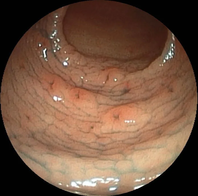 Endoscopy
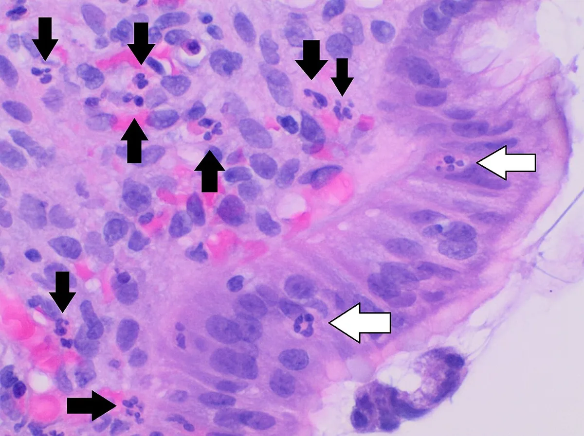 Histology
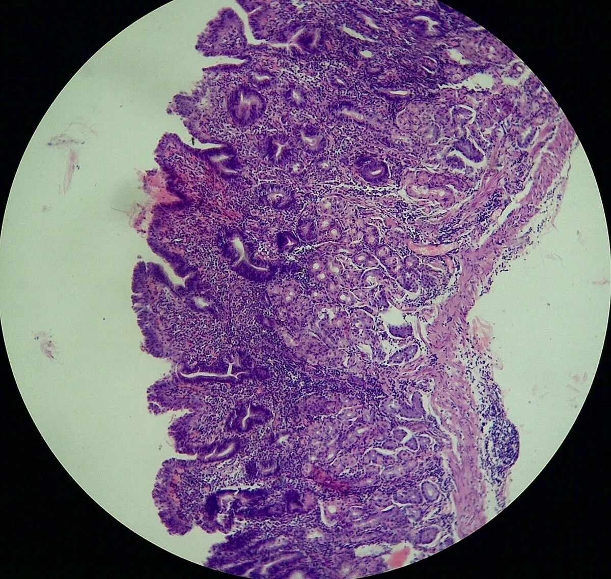 Histology
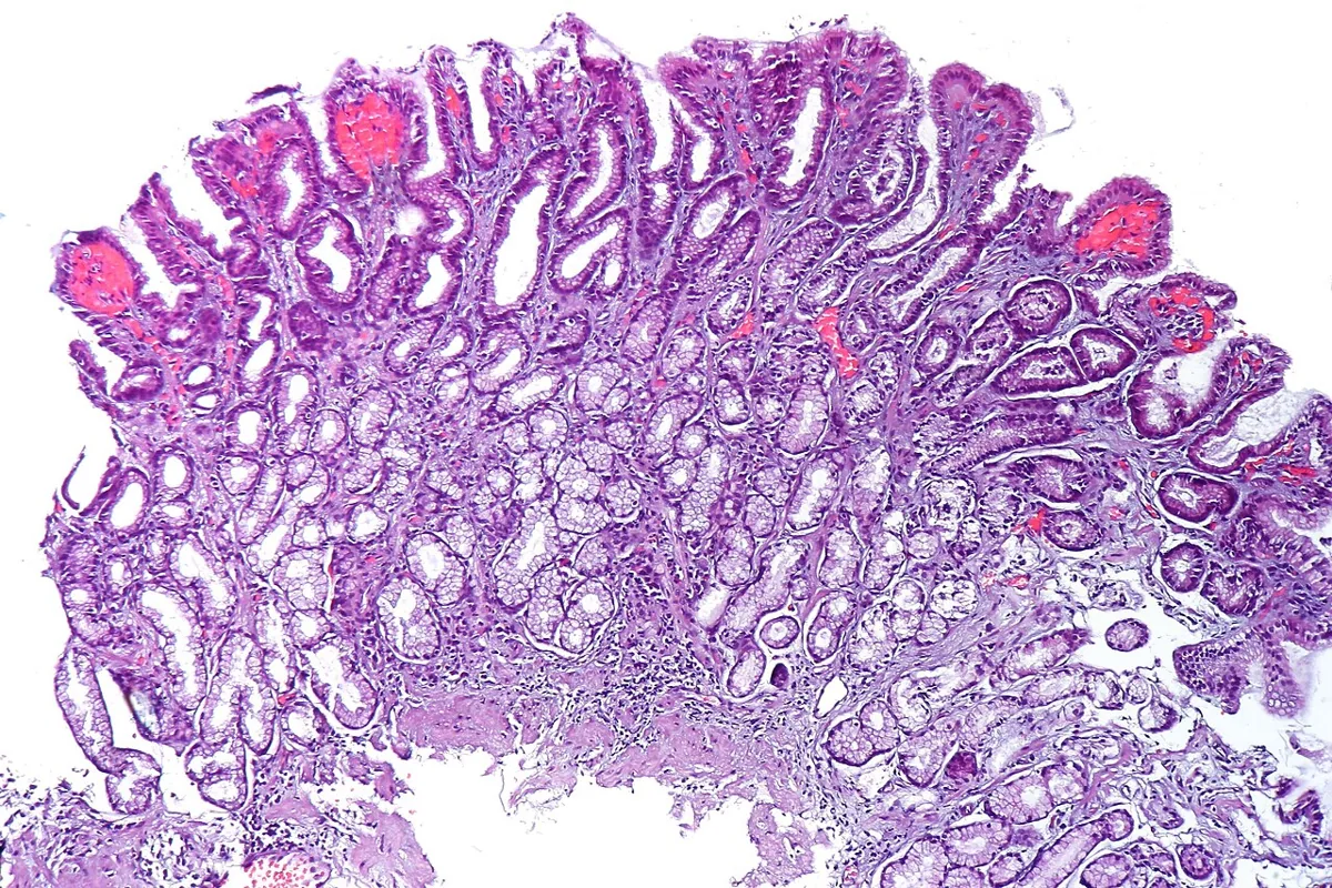 Histology
Definition
Gastritis is inflammation of the gastric mucosa. Causes include Helicobacter pylori infection, drugs such as NSAIDs, alcohol, stress and autoimmune processes. Many cases are asymptomatic; dyspepsia and gastrointestinal bleeding occur. In everyday use the term is often used loosely to non-specific upper abdominal complaints.
In German-speaking countries the chronic forms are classified etiologically as type A (autoimmune), type B (bacterial, H. pylori) and type C (chemical/reactive, e.g. due to NSAIDs or bile reflux).
Classification
- By severity of mucosal injury: erosive and non-erosive gastritis.
- By site: cardia, body, antrum.
- Histologically: acute (infiltration by neutrophils in the antrum and body) or chronic (with atrophy or metaplasia).
- Chronic antrum-predominant: loss of G cells with reduced gastrin secretion; often after long-standing H. pylori gastritis (type B).
- Chronic corpus-predominant: loss of oxyntic glands with reduced production of acid, pepsin and intrinsic factor; with autoantibodies to parietal cells, type A gastritis.
Aetiopathogenesis
- Erosive gastritis: commonly due to NSAIDs, alcohol or stress; less often due to damage from ionising rays, viral infection (e.g. cytomegalovirus), vascular injury, direct trauma (e.g. nasogastric tubes) or Crohn's disease. Superficial erosions may develop as soon as 12 hours after the injury.
- Acute stress gastritis: in critically ill patients, probably due to mucosal hypoperfusion; with head injury or burns, also increased acid secretion.
- Non-erosive gastritis: mainly the result of H. pylori infection.
- Autoimmune metaplastic atrophic gastritis (type A): inherited as an autosomal dominant trait; antibodies against parietal cells and their components (intrinsic factor, H⁺/K⁺-ATPase) lead to hypochlorhydria and intrinsic factor deficiency. Hypochlorhydria causes G-cell hyperplasia with often markedly raised gastrin (frequently > 1000 pg/ml), which induces enterochromaffin-like cell hyperplasia and occasionally neuroendocrine tumors.
- Association: Hashimoto's thyroiditis; up to one third of people with autoimmune thyroid disease also have autoimmune gastritis.
Clinical features
- Mild erosive gastritis: often asymptomatic, sometimes dyspepsia, nausea or vomiting; the first sign is often hematemesis, melaena or blood in the nasogastric aspirate, usually 2–5 days after the inciting event. Bleeding is usually mild to moderate, but can be massive with deep ulceration, particularly in stress gastritis.
- H. pylori gastritis: usually asymptomatic, sometimes mild dyspepsia; deep gastritis is more likely to cause symptoms.
- Autoimmune gastritis: itself causes few, non-specific symptoms (upper abdominal discomfort); vitamin B12 deficiency with megaloblastic (pernicious) anemia, fatigue and weakness, and neurological deficits (subacute combined degeneration) with reduced position and vibration sense, weakness and hyporeflexia.
- Complications of type A gastritis: gastric adenocarcinoma (about threefold increased risk) and neuroendocrine tumors; intestinal metaplasia may lead to stomach cancer.
Histology
- Superficial gastritis: lymphocytes and plasma cells mixed with neutrophils, superficial in the antrum, body or both; usually without atrophy or metaplasia.
- Deep gastritis: mononuclear cells and neutrophils throughout the mucosa to the level of the muscularis, patchy distribution.
- Atrophy: loss of gastric glands; with complete atrophy, secretion of acid and pepsin falls and intrinsic factor may be lost.
- Mucous gland (pseudopyloric) metaplasia: replacement of glands by mucous glands in severe atrophy, especially along the lesser curvature.
- Intestinal metaplasia: usually begins in the antrum; goblet cells, endocrine cells and rudimentary villi; the incomplete form resembles colonic mucosa and frequently shows dysplasia.
Diagnosis
- Endoscopy: confirms erosive and non-erosive gastritis; non-erosive gastritis is often discovered incidentally. In atrophy, the mucosa may look normal for a long time; in advanced stages submucosal vessels become visible.
- H. pylori testing: once gastritis has been identified.
- Autoimmune gastritis: confirmation by endoscopic biopsy; measurement of vitamin B12; parietal cell antibodies are usually present.
- Laboratory tests: full blood count (anemia), raised gastrin in type A.
Keep learning in the app
Further reading (open access)
Cross-references
More topics: Gastroenterology
Note: Learning content for medical education – not a treatment recommendation and no substitute for diagnosis or treatment decisions in individual cases. Treatment and management are deliberately not covered on this page.