Gallstones and acute cholecystitis
- Synonyms
- gallstones, gallstone disease, biliary colic, gallbladder inflammation, cholelithiasis, cholecystitis
- Specialty
- Internal medicine · Gastroenterology
- Images
- Ultrasound 2 · CT 1 · Gross specimen 1
- Last updated
- 10/2026 · Dr. Pascal Bafteh
Contents
Images (4)
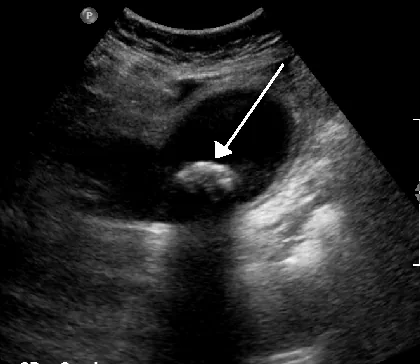 Ultrasound
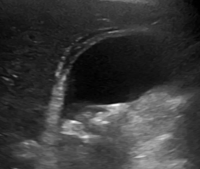 Ultrasound
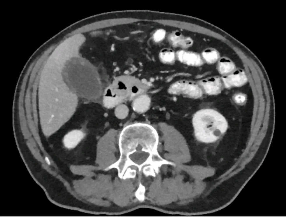 CT
CT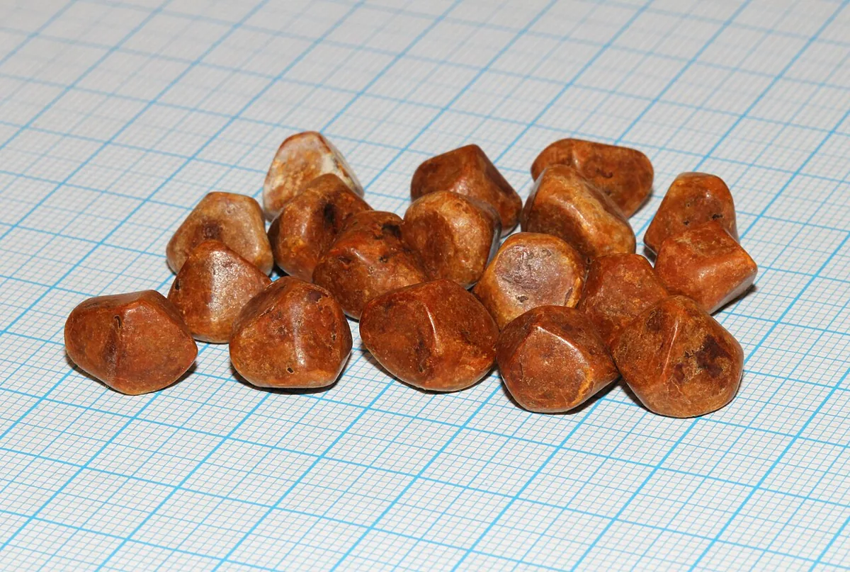 Gross specimen
Definition
Cholecystolithiasis (cholelithiasis in the narrower sense) is the presence of one or more gallstones in the gallbladder. Most disorders of the biliary tract result from gallstones. Gallstones are typically asymptomatic; the most common symptom is biliary colic.
Acute cholecystitis is inflammation of the gallbladder developing over hours, usually because a gallstone obstructs the cystic duct. It is the most common complication of cholelithiasis; at least 90% of patients have gallstones. Acalculous cholecystitis occurs without stones.
Classification
Stone types:
- Cholesterol stones: more than 85% of gallstones in Western countries
- Black pigment stones: small and hard, composed of calcium bilirubinate and inorganic calcium salts; favored by alcohol-related liver disease, chronic hemolysis and older age
- Brown pigment stones: soft and greasy, composed of bilirubinate and fatty acids; form during infection, inflammation and parasitic infestation (e.g. liver flukes) and arise in the bile ducts
Clinical forms: asymptomatic stones, biliary colic, acute cholecystitis, chronic cholecystitis (fibrotic, contracted gallbladder after recurrent inflammation) and complications such as choledocholithiasis, cholangitis and gallstone pancreatitis.
Occurrence & epidemiology
In the United States, an estimated 14% of people over 20 years have gallstones; prevalence doubled between 1988 and 2020. In some northern European countries it is as high as about 20%. Prevalence increases with age and plateaus in the sixth decade. Up to 80% of people with gallstones are asymptomatic. Acalculous cholecystitis accounts for only a small proportion of acute cholecystitis.
Aetiopathogenesis
Risk factors: female sex, obesity (especially in men), older age, physical inactivity, insulin resistance, lipid abnormalities, atherosclerotic heart disease and family history.
Stone formation: sludge (microlithiasis) composed of calcium bilirubinate, cholesterol microcrystals and mucin is often the precursor; it develops during gallbladder stasis, e.g. in pregnancy or with parenteral nutrition. A prerequisite for cholesterol stones is supersaturation of bile with cholesterol – usually due to excessive cholesterol secretion (e.g. in obesity or diabetes), less often due to decreased bile salt or lecithin secretion. Mucin accelerates precipitation of microcrystals, which aggregate and grow when gallbladder contractility is impaired. Gallstones grow about 1–2 mm per year and take 5–20 years before causing symptoms.
Acute cholecystitis: a stone impacted in the cystic duct causes bile stasis; inflammatory enzymes (e.g. phospholipase A, which converts lecithin to lysolecithin) and mediators such as prostaglandins damage the mucosa, which secretes more fluid. Distension worsens mucosal damage and ischemia; bacterial infection may supervene, with a risk of necrosis and perforation. Risk factors for acalculous cholecystitis are postoperative states and critical illness (burns, sepsis, shock, trauma), prolonged fasting or parenteral nutrition, immunodeficiency, vasculitis, diabetes and atherosclerosis.
Clinical features
Biliary colic: typically begins in the right upper quadrant but is often poorly localized (especially in older people and people with diabetes) and may radiate to the back or arm. The pain begins suddenly, becomes intense within 15 minutes to 1 hour, remains at a steady intensity (not colicky) usually for less than 6, occasionally up to 12 hours, and then subsides over 30–90 minutes. Nausea and vomiting are common, fever and chills are absent without cholecystitis. Between episodes, patients feel well. Gas, bloating and nausea are not specific gallbladder symptoms, and fatty food is not a specific trigger.
Acute cholecystitis: similar but more severe and longer-lasting pain (over 6 hours), frequent vomiting, right subcostal tenderness, spread of pain to the right scapula or upper back. Within a few hours a positive Murphy sign (inspiratory arrest on palpation of the right upper quadrant during deep inspiration) and guarding develop; usually low-grade fever. In older people, non-specific systemic symptoms may be the only signs. Colic lasting more than 12 hours, especially with vomiting or fever, suggests cholecystitis or pancreatitis.
Complications: empyema, gangrene and perforation (increasing pain, high fever, rigors, peritoneal signs); jaundice and acholic stool with bile duct involvement; Mirizzi syndrome (an impacted cystic duct stone narrows the common bile duct); gallstone pancreatitis; cholecystoenteric fistula with possible gallstone ileus. In critically ill patients, acalculous cholecystitis often shows only as abdominal distension or unexplained fever.
Diagnosis
- Abdominal ultrasound: the preferred test for gallbladder stones, sensitivity and specificity 95%; also detects sludge. In acute cholecystitis, tenderness over the gallbladder (sonographic Murphy sign), pericholecystic fluid and wall thickening
- CT and MRI: alternatives; show complications such as perforation, pancreatitis and duct stones, MRI with MRCP if choledocholithiasis is suspected
- Endoscopic ultrasound: also detects small stones under 3 mm
- X-ray: only about 10–15% of stones are calcified and visible
- Cholescintigraphy: if ultrasound is equivocal; failure of the gallbladder to fill indicates an obstructed cystic duct; the most reliable test in acalculous cholecystitis
- Laboratory tests: usually normal with uncomplicated stones; in acute cholecystitis frequent leukocytosis with left shift, liver tests normal or slightly elevated, mild cholestasis (bilirubin up to 4 mg/dl) common; more marked increases, especially with lipase above three times normal, suggest bile duct obstruction
Keep learning in the app
Further reading (open access)
Cross-references
More topics: Gastroenterology
Note: Learning content for medical education – not a treatment recommendation and no substitute for diagnosis or treatment decisions in individual cases. Treatment and management are deliberately not covered on this page.