Clostridioides difficile infection
Board exam relevance: in 2 of 105 exam reports · rank 142- Synonyms
- C. diff, C. difficile, Clostridium difficile, CDI, pseudomembranous colitis, antibiotic-associated colitis
- Specialty
- Internal medicine · Gastroenterology
- Images
- Endoscopy 1 · CT 1 · Histology 1 · Gross specimen 1
- Last updated
- 10/2026 · Dr. Pascal Bafteh
Contents
Images (4)
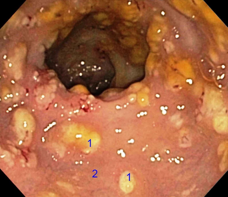 Endoscopy
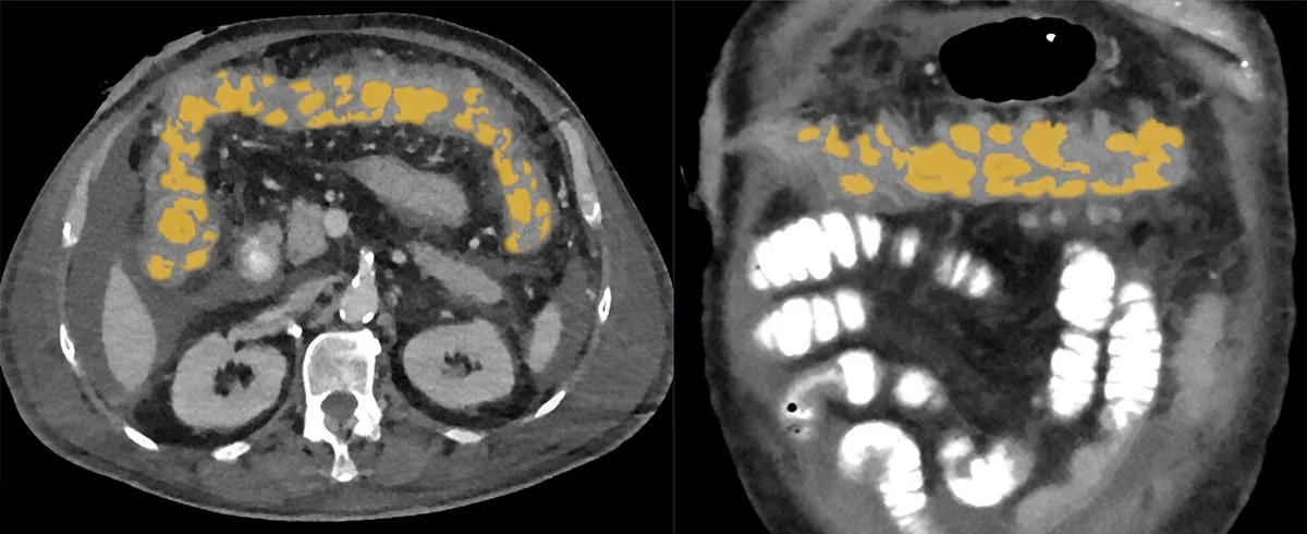 CT
CT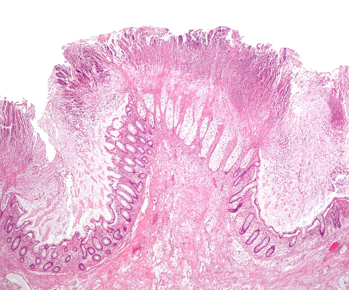 Histology
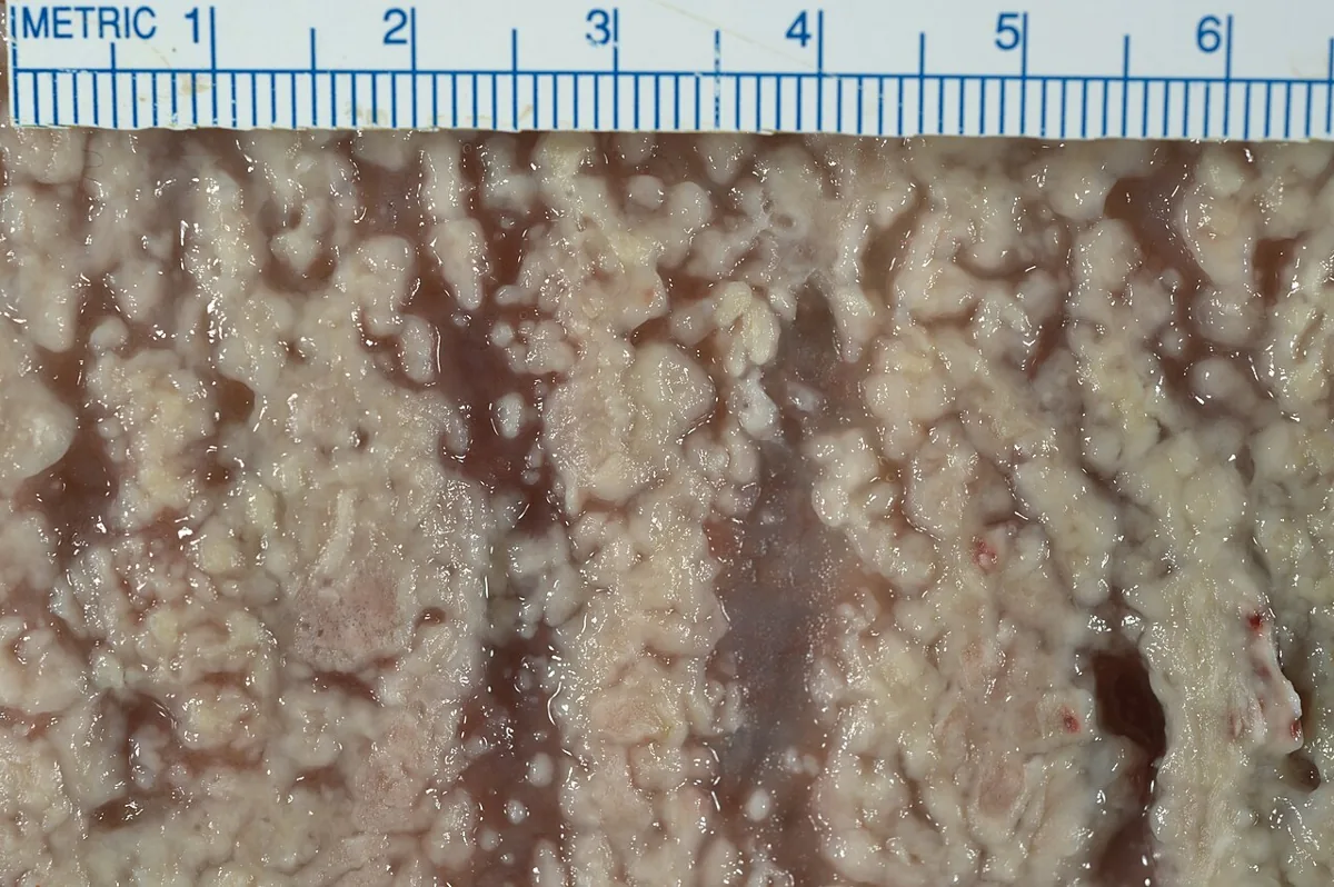 Gross specimen
Definition
Clostridioides difficile infection (CDI) is a toxin-mediated intestinal infection. Toxins of C. difficile cause pseudomembranous colitis, typically after intake of antibacterial drugs. C. difficile is the most common cause of antibiotic-associated colitis.
According to the case definition of the Robert Koch Institute, CDI is present in diarrhea or toxic megacolon together with detection of toxin A and/or B or toxin-producing C. difficile in the stool, in endoscopically proven pseudomembranous colitis, or with histopathological evidence.
Classification
- By place of acquisition: healthcare-associated or community-acquired; community-acquired CDI has symptom onset before or on the day of hospital presentation without a stay in a healthcare facility during the preceding twelve weeks
- Fulminant colitis: severe acute inflammation of the colon with systemic toxicity
- Relapse: a new episode within two months after improvement of a previous episode; if more than two months have passed, the episodes are considered separate events
Occurrence & epidemiology
C. difficile is common in the environment (soil, water, household pets). Asymptomatic colonization is relatively uncommon in healthy adults and considerably more frequent in patients in hospital and residents of long-term care facilities; in young children the organizm is frequently detected (up to 80%). C. difficile causes about 15–20% of antibiotic-associated diarrhea and more than 95% of pseudomembranous colitis.
Healthcare-associated cases are declining, whereas community-associated cases are slowly increasing. The more virulent strain BI/NAP1/027 (ribotype 027) produces substantially more toxin, causes more severe illness with more frequent relapses and is more transmissible; it is prominent in hospital outbreaks. Recurrences occur in 15–20% of patients, usually within a few weeks.
Aetiopathogenesis
Pathogen
- Pathogen: C. difficile — gram-positive spore-forming anaerobe.
- Pathogenicity factors: toxin A (enterotoxin) + toxin B (cytotoxin); hypervirulent ribotype 027 also produces binary toxin.
Disease develops through overgrowth of endogenous C. difficile in the gut or through infection from an external source. The spores are tolerant of heat, desiccation and many chemical agents; transmission occurs by oral ingestion, directly through contact, via the hands of staff or contaminated surfaces. The key predisposing factor is alteration of the gut flora by antibacterial drugs. In principle almost any drug class can be involved, with a particularly high risk after cephalosporins (especially third-generation), clindamycin and other broad-spectrum agents. Certain anticancer drugs can also trigger CDI. Not all strains produce toxins; some people carry toxin-producing strains without symptoms, especially infants.
Further risk factors:
- older age, severe underlying disease
- prolonged hospital stay, living in a nursing home
- drugs that lower gastric acid (risk increased about 2- to 3-fold)
- postoperative state after abdominal operations
Clinical features
- Typical: watery diarrhea, abdominal pain, fever, leukocytosis
- Severe CDI (criteria according to IDSA/SHEA 2017): white blood cell count of at least 15,000/µL or serum creatinine above 1.5 mg/dL
- Fulminant CDI: hypotension or shock, ileus, megacolon
Symptoms typically begin 5–10 days after starting antibacterial drugs but may occur on the first day or up to two months later. Diarrhea ranges from mild and semiformed to frequent and watery with a characteristic foul odor, and is occasionally bloody. Cramping and abdominal pain are common, nausea and vomiting rare; the abdomen is slightly tender. Fever, leukocytosis and early hypoalbuminemia due to massive protein loss may occur.
In fulminant colitis, patients appear very ill, with more pain, tachycardia and a distended, tender abdomen. Rarely, diarrhea is absent because of paralytic ileus. Complications include toxic megacolon, colonic perforation with peritoneal signs, sepsis and acute abdomen; reactive arthritis occurs rarely.
Histology
The toxins act mainly on the colon: it secretes fluid and forms characteristic pseudomembranes – discrete, yellowish-white, easily dislodged plaques that may coalesce in severe cases.
Diagnosis
- Suspicion: new, persistent diarrhea within two months of antibacterial drug use or 72 hours after entering hospital without another explanation (e.g. laxatives, tube feeding)
- Testing in new-onset, unexplained diarrhea with at least 3 unformed stools in 24 hours, as colonization is common; only unformed stool
- Glutamate dehydrogenase (GDH) antigen: produced by all strains; sensitive and rapid but says nothing about toxin production
- Toxin assay (ELISA): specific for active disease but not very sensitive
- Nucleic acid amplification (PCR) for the toxin gene: very sensitive for toxigenic strains but does not show whether toxin is currently produced; often remains positive after the illness has resolved
- Stepwise testing: GDH and toxin assays; if both agree, disease is considered confirmed or excluded, and discordant results are resolved by PCR
- Culture: the most sensitive detection method, basis for typing
- Sigmoidoscopy in ileus or nondiagnostic toxin assays (pseudomembranes); X-ray and CT if fulminant colitis, perforation or megacolon is suspected
Keep learning in the app
Further reading (open access)
Cross-references
More topics: Gastroenterology
Note: Learning content for medical education – not a treatment recommendation and no substitute for diagnosis or treatment decisions in individual cases. Treatment and management are deliberately not covered on this page.