Incontinentia pigmenti
- Synonyms
- Bloch-Sulzberger syndrome
- Specialty
- Dermatology · Hereditary skin diseases
- Images
- Clinical 2
- In the app
- 3 flashcards
- Last updated
- 10/2026 · Dr. Pascal Bafteh
Contents
Images (2)
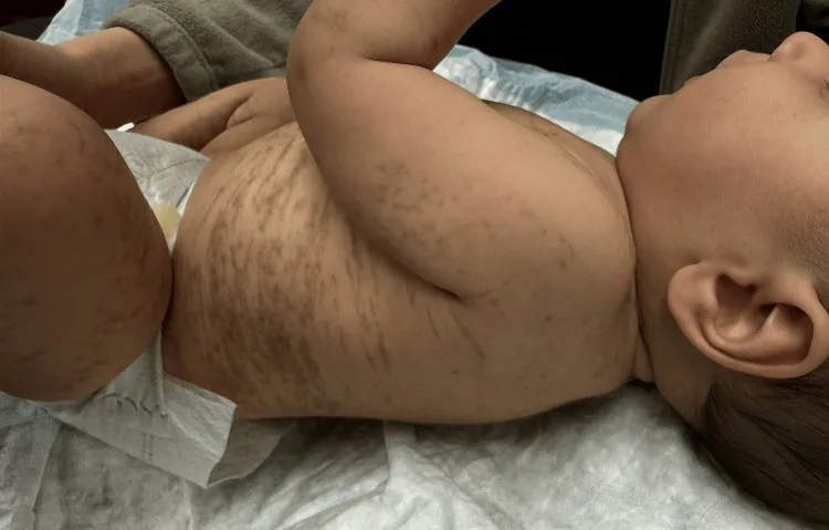
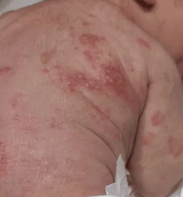
Definition
- Genetics: X-dominant, NEMO mutation — lethal in utero in male fetuses.
- 4 Stages (Blaschko-linear): bullous → verrucous → hyperpigmented → hypopigmented.
Clinical features
- Stage 1 (vesicular): in about 90% at birth or within the first 2 weeks of life, red blisters arranged in lines along Blaschko's lines, mostly on the limbs and scalp; lasting weeks to months, with possible recurrences in the first months of life.
- Stage 2 (verrucous): warty or pustular lesions with thick crusts, usually for months, rarely longer than a year.
- Stage 3 (hyperpigmented): whorled, blue-grey to brown pigmentation, usually appearing in the first months of life and slowly fading with age.
- Stage 4 (atrophic-hypopigmented): pale, hairless, scar-like patches or streaks that develop in adolescence and persist into adulthood (in 30–75%).
- In addition dental anomalies (over 80%; missing, peg-shaped teeth), hair and nail changes, eye involvement (20–35%, abnormal retinal vessel growth) and neurological involvement (up to 30%, mainly seizures).
Diagnosis
- Clinical picture with the typical sequence of stages; histology in the vesicular stage shows eosinophilic spongiosis.
- Molecular detection of an IKBKG (NEMO) mutation; ophthalmological examination in infancy because of possible retinal vascular changes.
Differential diagnoses
 Epidermoid cyst
 Aplasia cutis congenita
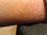 Ichthyosis
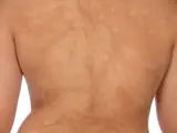 Hypomelanosis of Ito (incontinentia pigmenti achromians)
- Blaschko's lines
- Lichen striatus
- Naevus depigmentosus (achromic naevus)
Image credits for the thumbnails
- Epidermoid cyst: unknown (Commons user), “Epidermal Cyst ear”, CC BY-SA 3.0, via Wikimedia Commons · CC BY-SA 3.0 · Source
- Aplasia cutis congenita: Liwen Xu, Kevin X. Liu, Maryanne M. Senna, CC BY 4.0, via Wikimedia Commons · CC BY 4.0 · Source
- Ichthyosis: Skoch3, “2012 April mosaic lines left calf”, CC BY-SA 3.0, via Wikimedia Commons · CC BY-SA 3.0 · Source
- Hypomelanosis of Ito (incontinentia pigmenti achromians): Trägårdh M, Thomsen CR, Thorninger R et al. (J Med Case Rep 2014), “Hypomelanosis of Ito presenting with pediatric orthopedic issues: a case report”, CC BY 2.0, via PubMed Central (PMC4077238) · CC BY 2.0 · Source
Treatment principles
- There is no causal treatment; the skin lesions usually pass through their stages spontaneously and need at most symptomatic local care, such as protection against infection in the vesicular stage.
- Interdisciplinary care is crucial, particularly early and regular ophthalmological examinations because of retinal vasculopathy, together with neurological and dental follow-up.
- Genetic counselling is part of management.
Specific treatment regimens with drugs and doses are covered in the DermaFuchs app.
Keep learning in the app
References (selection)
Cross-references
Mentioned in
Keep learning
Keep learning with flashcards, exam questions and spot diagnoses in the DermaFuchs app – free, in your browser or as an app.
Related pages: Epidermoid cyst · Aplasia cutis congenita · Ichthyosis
Note: This page is intended for medical education and does not replace diagnosis or treatment decisions in individual cases.