Milia (milium)
- Synonyms
- Milium, milia, milk spots
- Specialty
- Dermatology · Skin appendages
- Images
- Clinical 2
- In the app
- 1 flashcard
- Last updated
- 10/2026 · Dr. Pascal Bafteh
Contents
Images (2)
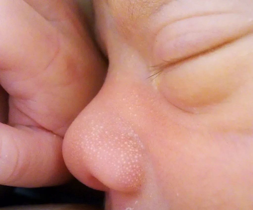
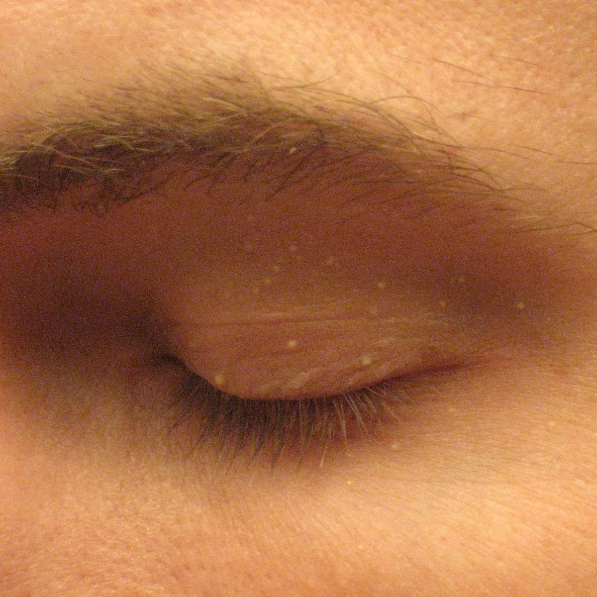
Definition
- Definition: 1–2 mm whitish-yellow, firm epidermal keratin cysts — essentially a miniature epidermal cyst.
- Primary milia: no preceding damage, predilection periorbital and cheeks. As milia neonatorum in about 40–50 % of newborns and self-limiting.
- Distinction: syringoma (skin-coloured to yellowish dermal papules, lower lid), xanthelasma (yellow plaques), sebaceous hyperplasia (central dell) — milia are firm and white, without a central dell.
Aetiopathogenesis
Primary milia arise spontaneously from vellus hair follicles. Secondary (traumatic) milia form from eccrine sweat ducts at sites of skin damage, for example in blistering diseases (porphyria cutanea tarda, epidermolysis bullosa, bullous pemphigoid), after burns or dermabrasion.
Histology
Small cysts filled with laminated keratin and lined by stratified squamous epithelium with a granular layer – histologically a miniature epidermoid cyst.
Differential diagnoses

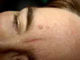
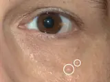
Image credits for the thumbnails
- Epidermoid cyst: unknown (Commons user), “Epidermal Cyst ear”, CC BY-SA 3.0, via Wikimedia Commons · CC BY-SA 3.0 · Source
- Hidrocystoma (eccrine and apocrine): unknown (Commons user), “Eccrine hydrocystoma 01”, CC BY 2.0, via Wikimedia Commons · CC BY 2.0 · Source
- Trichoepithelioma: Klaus D. Peter, Wiehl, Germany, “Trichoepithelioma”, CC BY 3.0 de, via Wikimedia Commons · CC BY 3.0 de · Source
- Sebaceous hyperplasia: Klaus D. Peter, Wiehl, “Seborrhoeic dermatitis2” (Commons label differs from the diagnosis), CC BY 3.0 DE, via Wikimedia Commons · CC BY 3.0 DE · Source
- Syringoma: Mark A. Taff, “Syringoma”, CC BY-SA 3.0 US, via Wikimedia Commons · CC BY-SA 3.0 US · Source
Treatment principles
- Milia are harmless; in neonates they regress spontaneously, so no treatment is required.
- For cosmetic reasons, they can be expressed after opening the overlying epidermis with a lancet or needle, or removed by electrocautery or ablative laser.
- Topical retinoids may be considered for multiple milia; in secondary milia, underlying conditions such as bullous dermatoses should be taken into account.
Specific treatment regimens with drugs and doses are covered in the DermaFuchs app.
Keep learning in the app
References (selection)
Cross-references
Mentioned in
More diseases of the skin appendages
Keep learning: test yourself
One question from the DermaFuchs app on Milia (milium) – tap an answer.
Question 1 of 1
Where do primary milia commonly appear without preceding damage?
Answer
Correct: periorbital and cheeks. Primary milia have a predilection for the periorbital area and cheeks, often appearing in newborns.
More questions like these – with explanations and spaced repetition – in the app.
Note: This page is intended for medical education and does not replace diagnosis or treatment decisions in individual cases.