Epidermolysis bullosa acquisita
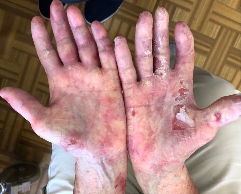
Fundamentals & Histology
- Definition: Rare, acquired chronic autoimmune blistering dermatosis of the basement membrane.
- Pathogenesis: IgG autoantibodies against Collagen VII, which destroy the anchoring fibrils beneath the lamina densa.
- Histology: Characteristic is a subepidermal cleft formation with variably pronounced inflammatory infiltrate (mechanobullous vs. inflammatory type).
- Clinical Features: Mechanically induced blisters at friction points, which heal with scarring and milia formation.
Diagnostics
- Direct Immunofluorescence (DIF): Detection of linear IgG and C3 deposits along the dermo-epidermal junction zone.
- Differentiation (Salt-split-skin): In EBA, immunoglobulins bind to the dermal side (blister floor), in contrast to bullous pemphigoid.
- Serology: Detection of anti-collagen VII antibodies by ELISA or indirect immunofluorescence on monkey esophagus.
Differential diagnoses
- Pemphigus vulgaris
- Linear IgA dermatosis
- Generalized pustular psoriasis (von Zumbusch)
- Bullous pemphigoid
- Hereditary epidermolysis bullosa
- Pemphigoid gestationis
Practise Epidermolysis bullosa acquisita in the app
Flashcards with spaced repetition, exam questions and spot-the-diagnosis on this topic – in DermaFuchs, free of charge.
In the DermaFuchs app: 3 flashcards · 2 clinical images
Open in browser Download on the App StoreSources (selection)
More blistering diseases
Note: This page is intended for medical education and does not replace diagnosis or treatment decisions in individual cases. Treatment and follow-up content is available in the app.