Erythema dyschromicum perstans (ashy dermatosis)
- Synonyms
- Ashy dermatosis, Dermatosis cenicienta, aschgraue Dermatose, Erythema dyschromicum perstans, ashy dermatosis of Ramirez, EDP
- Specialty
- Dermatology · Pigmentation disorders
- Images
- Clinical 1
- In the app
- 3 flashcards
- Last updated
- 10/2026 · Dr. Pascal Bafteh
Contents
Images (1)
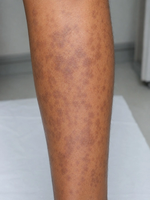 AI illustration
Definition
Erythema dyschromicum perstans (ashy dermatosis of Ramírez) is an acquired dermal macular hyperpigmentation with well-circumscribed, round to oval or irregular grey patches on the face, neck and trunk. Because of its histological similarity it is often classified as a variant of lichen planus pigmentosus; the terms for these acquired macular pigmentations are used inconsistently worldwide, and an international consensus statement addresses the terminology.
Occurrence & epidemiology
It mainly affects people with darker skin, most frequently from Latin America and India; cases in lighter skin have been reported. It can occur at any age but is more frequent in young adults; women are affected more often than men.
Aetiopathogenesis
The cause is unknown. Proposed factors include genetic susceptibility, contact allergy to cosmetics and hair dye, chemicals (ammonium nitrate, barium sulphate), whipworm infestation, viral infections and drugs.
Clinical features
- Ash-grey patches, distributed symmetrically or unilaterally; no symptoms.
- Early lesions may be reddish, with a more pronounced, slightly raised border – this phase is not always observed.
- Patients are otherwise well, with no associated disease or blood test abnormality.
- The patches often persist unchanged for years; some clear spontaneously.
Histology
Early lesions show mild vacuolar degeneration of the basal layer; established patches show pigmentary incontinence with dermal melanophages.
Diagnosis
- Often a clinical diagnosis; a skin biopsy supports it.
- Dermoscopy: in one series (60 lesions in 15 patients) the most common findings were irregular linear dots and globules on a pinkish-brown background (mainly in early lesions) and irregular linear vessels.
Differential diagnoses
- Ashy dermatosis (= Erythema dyschromicum perstans)
- Lupus erythematosus
- Pityriasis rosea
- Psoriasis vulgaris (plaque psoriasis)
- Erythema ab igne
- Fixed drug eruption (FDE)
- Addison disease
- Schamberg's disease (progressive pigmented purpura)
Keep learning in the app
References (selection)
- DermNet: Erythema dyschromicum perstans
- PubMed: A global consensus statement on ashy dermatosis, erythema dyschromicum perstans, lichen planus pigmentosus, idiopathic eruptive macular pigmentation, and Riehl's melanosis (PMID 30176055)
- PubMed: Dermoscopic Diagnosis of Ashy Dermatosis: A Retrospective Study (PMID 31807441)
Cross-references
Mentioned in
Note: This page is intended for medical education and does not replace diagnosis or treatment decisions in individual cases.