Paronychia
- Synonyms
- Nagelbettentzündung, Nagelumlauf, Umlauf, Nagelwallentzündung, Panaritium parunguale, nail fold infection
- Specialty
- Dermatology · Skin appendages
- Images
- Clinical 2
- In the app
- 5 flashcards
- Last updated
- 10/2026 · Dr. Pascal Bafteh
Contents
Images (2)
Definition
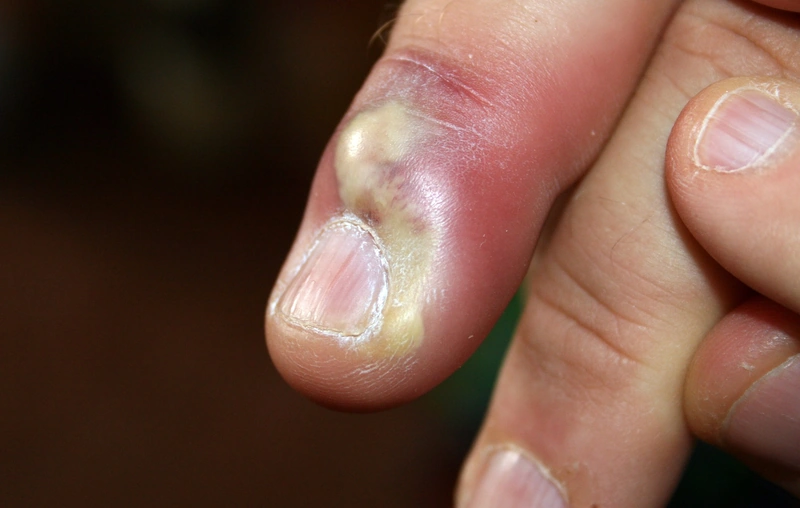
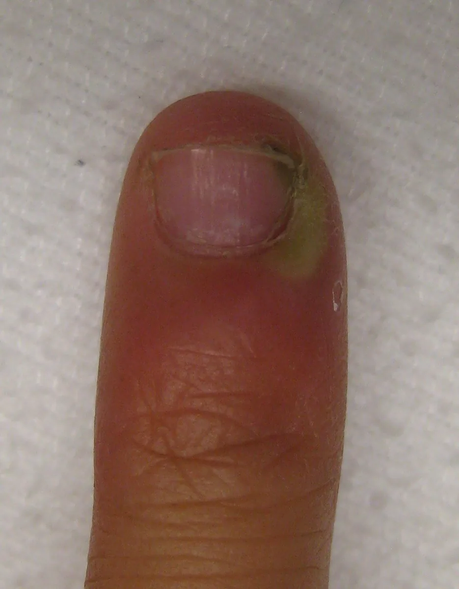
Paronychia is inflammation of the nail fold. Acute, usually bacterial paronychia is distinguished from chronic paronychia, which lasts more than six weeks and is mostly caused by moisture and irritation.
Aetiopathogenesis
- Acute: entry of bacteria (usually Staphylococcus aureus, streptococci) through minor injuries, hangnails, nail biting or nail-care procedures; herpes simplex can also cause paronychia (herpetic whitlow).
- Chronic: constantly cold, wet hands (e.g. cleaning, bar work, dairy farming, fishing), hand dermatitis or dermatitis of the nail fold; the nail fold lifts off the nail plate, allowing entry of organisms and irritants; Candida albicans and Pseudomonas are often cultured.
Clinical features
- Acute: rapidly developing redness, swelling and throbbing pain at the nail fold, often with pus collecting under the fold.
- Chronic: swollen, red, tender nail fold without cuticle, several fingers; transverse ridges and discoloration of the nail plate.
Diagnosis
- Clinical diagnosis; swab with pus, mycology in chronic courses, HSV PCR with vesicles.
- With single long-standing changes on one finger, exclusion of tumours (subungual melanoma, squamous cell carcinoma, metastases), especially with an atypical picture or lack of improvement.
Differential diagnoses
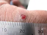
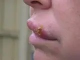
Image credits for the thumbnails
- Onychomycosis (nail fungus): Bobjgalindo, “Onychomycosis”, CC BY-SA 4.0, via Wikimedia Commons · CC BY-SA 4.0 · Source
- Nail psoriasis: Seenms, “Luszczyca paznokcia”, CC BY-SA 3.0, via Wikimedia Commons · CC BY-SA 3.0 · Source
- Pyogenic granuloma: kilbad, “Pyogenic granuloma on a finger-1”, CC BY 3.0, via Wikimedia Commons · CC BY 3.0 · Source
- Herpes simplex (cold sores): unknown (Commons user), “Cold sore”, Public Domain, via Wikimedia Commons · Public Domain · Source
Treatment principles
- Acute paronychia is treated with antiseptic soaks or compresses and topical antiseptics or antibiotics; an abscess must be incised and drained.
- Spreading infection or systemic symptoms warrant systemic antibiotic therapy.
- Chronic paronychia is primarily an irritant inflammatory condition: avoidance of moisture and irritants and topical glucocorticoids are central, with antimycotics added for Candida colonisation.
Specific treatment regimens with drugs and doses are covered in the DermaFuchs app.
Keep learning in the app
References (selection)
Cross-references
Keep learning
Keep learning with flashcards, exam questions and spot diagnoses in the DermaFuchs app – free, in your browser or as an app.
Related pages: Onychomycosis (nail fungus) · Nail psoriasis · Pyogenic granuloma
Note: This page is intended for medical education and does not replace diagnosis or treatment decisions in individual cases.