Pemphigus herpetiformis
- Synonyms
- herpetiformer Pemphigus, Pemphigus mit eosinophiler Spongiose, herpetiform pemphigus, pemphigus with eosinophilic spongiosis
- Specialty
- Dermatology · Blistering diseases
- Images
- Clinical 1
- In the app
- 1 flashcards
- Last updated
- 10/2026 · Dr. Pascal Bafteh
Contents
Images (1)
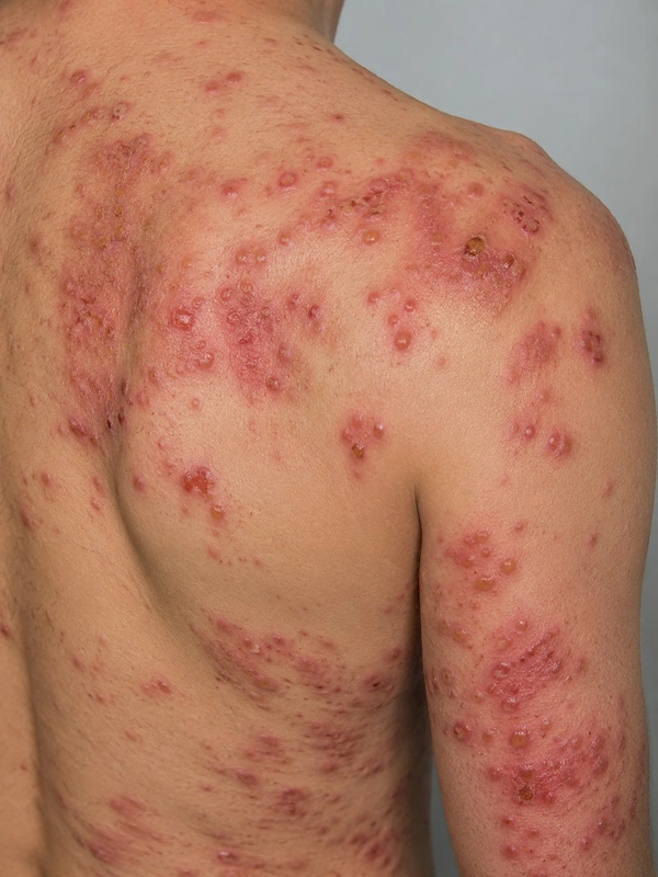 AI illustration
Definition
Pemphigus herpetiformis is a rare variant of pemphigus that combines the clinical picture of dermatitis herpetiformis with the immunological features of pemphigus; it was first described in 1975 by Jabłońska and colleagues. Clinically it may also resemble linear IgA bullous dermatosis or bullous pemphigoid.
Aetiopathogenesis
An autoimmune disease with autoantibodies mostly against desmoglein 1, less often against desmoglein 3; antibodies against desmocollins are increasingly being detected as well.
Clinical features
- Leading symptom is itch (present at onset in all 8 patients of one case series).
- Itchy, herpetiform grouped, intact vesicles or blisters with or without erosions and/or itchy annular or urticarial erythematous plaques.
- Some patients have peripheral eosinophilia (3 of 8 in the case series).
Histology
Intraepidermal eosinophils and/or neutrophils (eosinophilic spongiosis) and/or an intraepidermal split with or without acantholysis. In one case series all patients showed eosinophilic spongiosis, but only half showed acantholysis.
Diagnosis
- Direct immunofluorescence: intercellular deposits of IgG (with or without C3) in the epidermis.
- Serology: indirect immunofluorescence with IgG against the epithelial cell surface; autoantibodies against desmogleins (1, 3) and/or desmocollins (1–3).
- Proposed diagnostic criteria (literature review of 158 cases): one clinical, one histological and one immunological feature.
- The clinical and histological picture changes over time, so the diagnosis is often delayed.
Differential diagnoses
Keep learning in the app
References (selection)
- PubMed: Pemphigus herpetiformis: from first description until now (PMID 24472428)
- PubMed: Clinical, pathologic, and immunologic features of pemphigus herpetiformis: a literature review and proposed diagnostic criteria (PMID 30900757)
- PubMed: Pemphigus herpetiformis: a case series and review of the literature (PMID 25600350)
Cross-references
Note: This page is intended for medical education and does not replace diagnosis or treatment decisions in individual cases.