Lichen planus pemphigoides
- Synonyms
- Lichen ruber pemphigoides, LPP
- Specialty
- Dermatology · Blistering diseases
- Images
- Histology 1
- Last updated
- 10/2026 · Dr. Pascal Bafteh
Contents
Images (1)
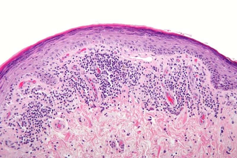 Histology
Definition
Lichen planus pemphigoides (LPP) is a very rare autoimmune subepidermal blistering dermatosis in which lichenoid skin lesions and tense blisters occur together with autoantibodies against type XVII collagen (BP180). It is now regarded as a distinct entity.
Occurrence & epidemiology
Its prevalence is estimated at about 1 per 1,000,000. The mean age at onset is about 46 years (range 4–85 years), well below that of bullous pemphigoid; children and adolescents are also affected. There is no clear sex predilection.
Aetiopathogenesis
The autoantibodies are mostly directed against the NC16A domain of type XVII collagen. Because the blisters almost always develop after the lichenoid lesions, it is thought that the lichenoid inflammation triggers the autoimmune response against basement membrane zone proteins (epitope spreading). Cases after drugs and viral infections (e.g. varicella, hepatitis B) have been described.
Clinical features
- Two primary lesions: itchy, violaceous, polygonal, shiny papules and plaques of lichen planus and tense blisters and erosions.
- The blisters typically develop after the lichenoid lesions and classically on previously unaffected skin (in bullous lichen planus, by contrast, on the lichen plaques).
- Mainly on the limbs; the course is usually milder and shorter than in bullous pemphigoid.
- Mucosal involvement with whitish striae is possible, as are purely mucosal forms; nail involvement up to loss of the nail plate.
Histology
- Blister: subepidermal separation with numerous eosinophils, as in bullous pemphigoid.
- Lichenoid lesion: picture of lichen planus with hypergranulosis, band-like lymphocytic infiltrate and Civatte bodies.
Diagnosis
- Direct immunofluorescence of a perilesional biopsy: deposits of IgG and C3 at the dermal-epidermal junction.
- Serology: circulating autoantibodies against type XVII collagen (e.g. ELISA; binding to the blister roof on salt-split skin).
- The diagnosis is confirmed by the typical clinical picture plus detection of autoantibodies; these antibodies are absent in bullous lichen planus.
Differential diagnoses
Keep learning in the app
References (selection)
Cross-references
Note: This page is intended for medical education and does not replace diagnosis or treatment decisions in individual cases.