Mucous membrane pemphigoid (cicatricial pemphigoid)
Exam relevance: in 2 of more than 50 board exam reports · rank 69
- Synonyms
- vernarbendes Pemphigoid, okuläres Pemphigoid, MMP, Schleimhaut-Pemphigoid, benignes Schleimhautpemphigoid, mucous membrane pemphigoid, cicatricial pemphigoid
- Specialty
- Dermatology · Blistering diseases
- Images
- Clinical 3
- In the app
- 1 flashcard
- Last updated
- 10/2026 · Dr. Pascal Bafteh
Contents
Images (3)
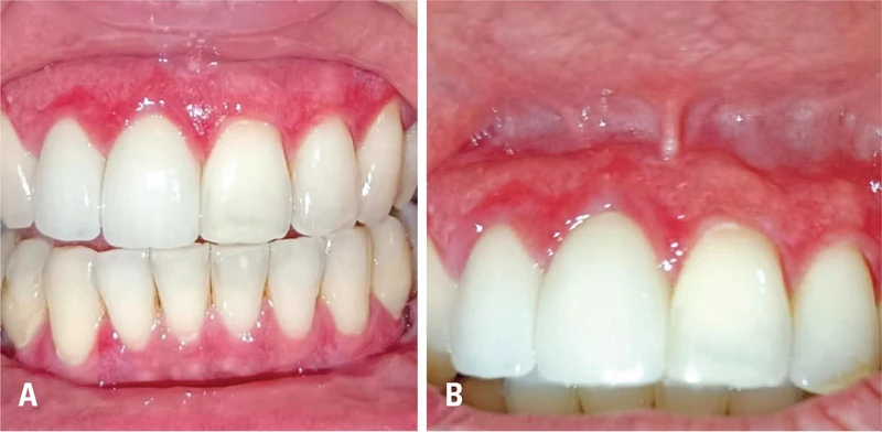
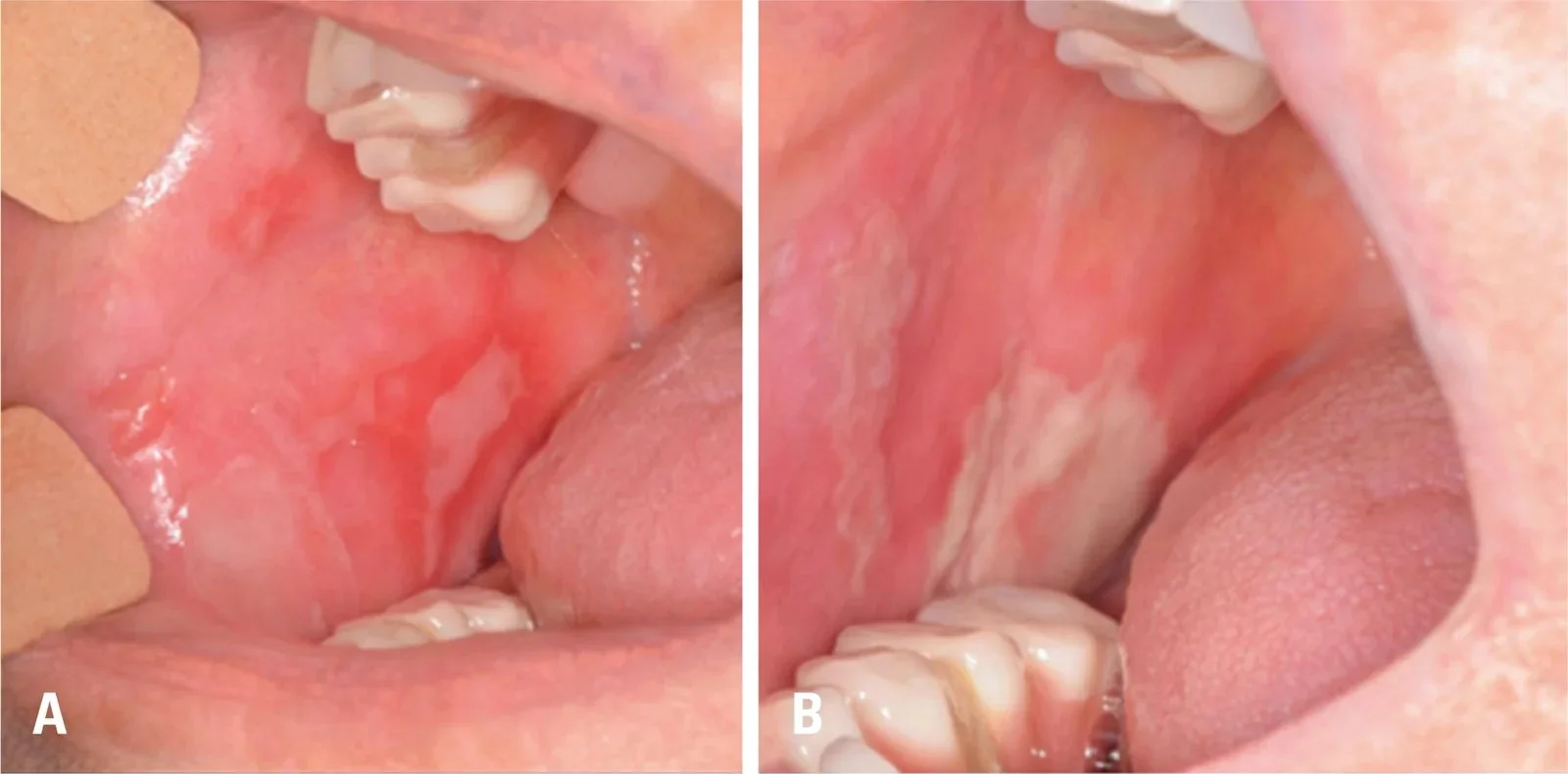
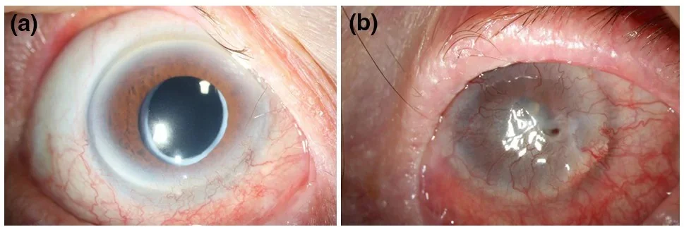
Definition
Mucous membrane pemphigoid (formerly cicatricial pemphigoid) is a group of chronic subepithelial autoimmune blistering diseases that predominantly affect the mucous membranes and tend to scar. The autoantibodies target structural proteins of the basement membrane zone: collagen XVII, BP230, laminin 332, integrin α6β4 and collagen VII.
Occurrence & epidemiology
The disease is rare (about 1.3–2.0 cases per million people per year), affects women more often than men and is predominantly a disease of the elderly with a peak at around 70 years; childhood cases have been reported.
Clinical features
- Oral mucosa (over 85%, most common site): blisters, patches or erosions on the gums, palate, tongue, lips and buccal mucosa, with painful eating.
- Eyes (about 65%): chronic conjunctivitis, entropion, adhesions between eyelid and globe (symblepharon), progressive scarring up to blindness.
- Other mucosae: nose, pharynx, larynx (hoarseness, dyspnoea), oesophagus (dysphagia), anogenital region.
- Skin: in about 25–30%, blisters mainly on the face, neck and scalp; Brunsting-Perry variant with scarring plaques on the head.
Histology
Subepithelial blistering after migration of inflammatory cells into the upper lamina propria with degradation of the basement membrane; later scarring.
Diagnosis
- Direct immunofluorescence of perilesional mucosa or skin: linear deposition of IgG and/or C3, sometimes IgA, along the basement membrane zone.
- Serology: indirect immunofluorescence and screening for antibodies to laminin 332; antibodies are often present only in low titres and are not always detected.
- Examination of all potentially affected mucosae for risk stratification: high risk with involvement of the conjunctiva, larynx, oesophagus, trachea or genital mucosa, low risk with oral, nasal or skin involvement only.
Differential diagnoses
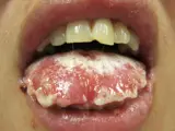
 Pemphigus vulgaris
 Stevens-Johnson syndrome
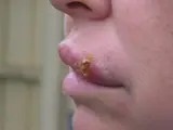 Herpes simplex (cold sores)
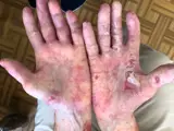 Epidermolysis bullosa acquisita
 Lichen planus
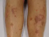
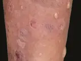 Lichen planus pemphigoides
Image credits for the thumbnails
- Pemphigus vulgaris: Mohammad2018, CC BY-SA 4.0, via Wikimedia Commons, cropped · CC BY-SA 4.0 · Source
- Stevens-Johnson syndrome: James Heilman, MD, “Mucosal desquamation in a person with Stevens–Johnson syndrome”, CC BY-SA 3.0, via Wikimedia Commons · CC BY-SA 3.0 · Source
- Herpes simplex (cold sores): unknown (Commons user), “Cold sore”, Public Domain, via Wikimedia Commons · Public Domain · Source
- Epidermolysis bullosa acquisita: Szymański, Kowalewski, Pietrzyk et al. (2023), “Case Report: Biological treatment of epidermolysis bullosa acquisita: report on four cases and literature review”, CC BY 4.0, via Wikimedia Commons · CC BY 4.0 · Source
- Lichen planus: James Heilman, MD, “Lichen Planus (2)”, CC BY-SA 3.0, via Wikimedia Commons · CC BY-SA 3.0 · Source
- Lichen planus pemphigoides: Gandhi V, Kumar P, Chauhan A (An Bras Dermatol 2022), CC BY 4.0, via PubMed Central (PMC9582882) · CC BY 4.0 · Source · cropped
Treatment principles
- Treatment depends on the risk of the sites involved: when only the oral mucosa or skin is affected, topical glucocorticoids and anti-inflammatory systemic agents are often sufficient.
- Involvement of the eyes, larynx, oesophagus or genitals requires early systemic immunosuppression because of the risk of scarring, with anti-CD20 antibodies or intravenous immunoglobulins in refractory disease.
- Care is multidisciplinary, involving ophthalmology and ENT among others.
Specific treatment regimens with drugs and doses are covered in the DermaFuchs app.
Keep learning in the app
References (selection)
Cross-references
Keep learning: test yourself
One question from the DermaFuchs app on Mucous membrane pemphigoid (cicatricial pemphigoid) – tap an answer.
Question 1 of 1
Which areas are predominantly affected in Mucous Membrane Pemphigoid?
Answer
Correct: oral mucosa & conjunctivae. Mucous Membrane Pemphigoid predominantly affects the oral mucosa and conjunctivae, with rare skin involvement.
More questions like these – with explanations and spaced repetition – in the app.
Note: This page is intended for medical education and does not replace diagnosis or treatment decisions in individual cases.