Bullous pemphigoid
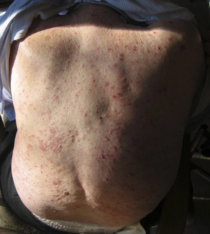
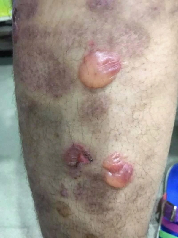
Overview
Autoantibodies: linear deposition along the basement membrane zone of IgG and C3 in a smooth, band-like pattern against hemidesmosomes or against BP180 & BP230. In histology, the salt-split skin method is used to demonstrate antibody deposition on the blister roof by achieving a separation at the lamina Lucida of the basement membrane through NaCl solution. This allows the disease to be distinguished from Epidermolysis Bullosa Acquisita (here antibodies at the blister floor). Triggers: Tumors, infections, UV radiation or medications such as DPP-4 inhibitors, ACE inhibitors, Rivaroxaban, Amoxicillin, Terbinafine.
Differential diagnoses
- Urticaria
- Pemphigus vulgaris
- Scabies
- Dermatitis herpetiformis (Duhring disease)
- TEN (toxic epidermal necrolysis)
- Epidermolysis bullosa acquisita
- Linear IgA dermatosis
- Pemphigoid gestationis
Practise Bullous pemphigoid in the app
Flashcards with spaced repetition, exam questions and spot-the-diagnosis on this topic – in DermaFuchs, free of charge.
In the DermaFuchs app: 4 flashcards · 8 clinical images
Open in browser Download on the App StoreSources (selection)
More blistering diseases
Note: This page is intended for medical education and does not replace diagnosis or treatment decisions in individual cases. Treatment and follow-up content is available in the app.