Lichen planopilaris
Overview
- Pathogenesis: Pathogenetically central is Th1/Th17 + CD8+ T-cells against bulge with IFN-γ apoptosis.
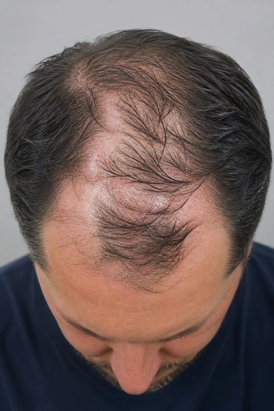
- Clinical features: circumscribed atrophic bald plaques with perifollicular erythema + perifollicular hyperkeratosis, pruritus, burning.
- Trichoscopy: Typical for LPP are perifollicular erythema + blue-gray dots — the blue-gray dots result from pigment incontinence after interface dermatitis at the bulge.
- Histology: Interface dermatitis at the bulge with lichenoid infiltrate + loss of sebaceous glands.
Differential diagnoses
- Folliculitis decalvans
- Alopecia areata
- Frontal fibrosing alopecia
- Discoid lupus erythematosus (CDLE)
- Psoriasis vulgaris (plaque psoriasis)
- Androgenetic Alopecia
- Lichen striatus
Practise Lichen planopilaris in the app
Flashcards with spaced repetition, exam questions and spot-the-diagnosis on this topic – in DermaFuchs, free of charge.
In the DermaFuchs app: 2 flashcards · 1 clinical images
Open in browser Download on the App StoreMore diseases of the skin appendages
- Acne fulminans
- Hidradenitis suppurativa (acne inversa)
- Acne vulgaris
- Alopecia areata
- Androgenetic alopecia
- Folliculitis (infectious)
- Milia (milium)
- Perioral dermatitis
Note: This page is intended for medical education and does not replace diagnosis or treatment decisions in individual cases. Treatment and follow-up content is available in the app.