Varicose veins
- Synonyms
- varices, spider veins, varicosis, bulging leg veins
- Specialty
- Internal medicine · Angiology
- Images
- Clinical 2 · Gross specimen 1 · X-ray 1
- Last updated
- 10/2026 · Dr. Pascal Bafteh
Contents
Images (4)
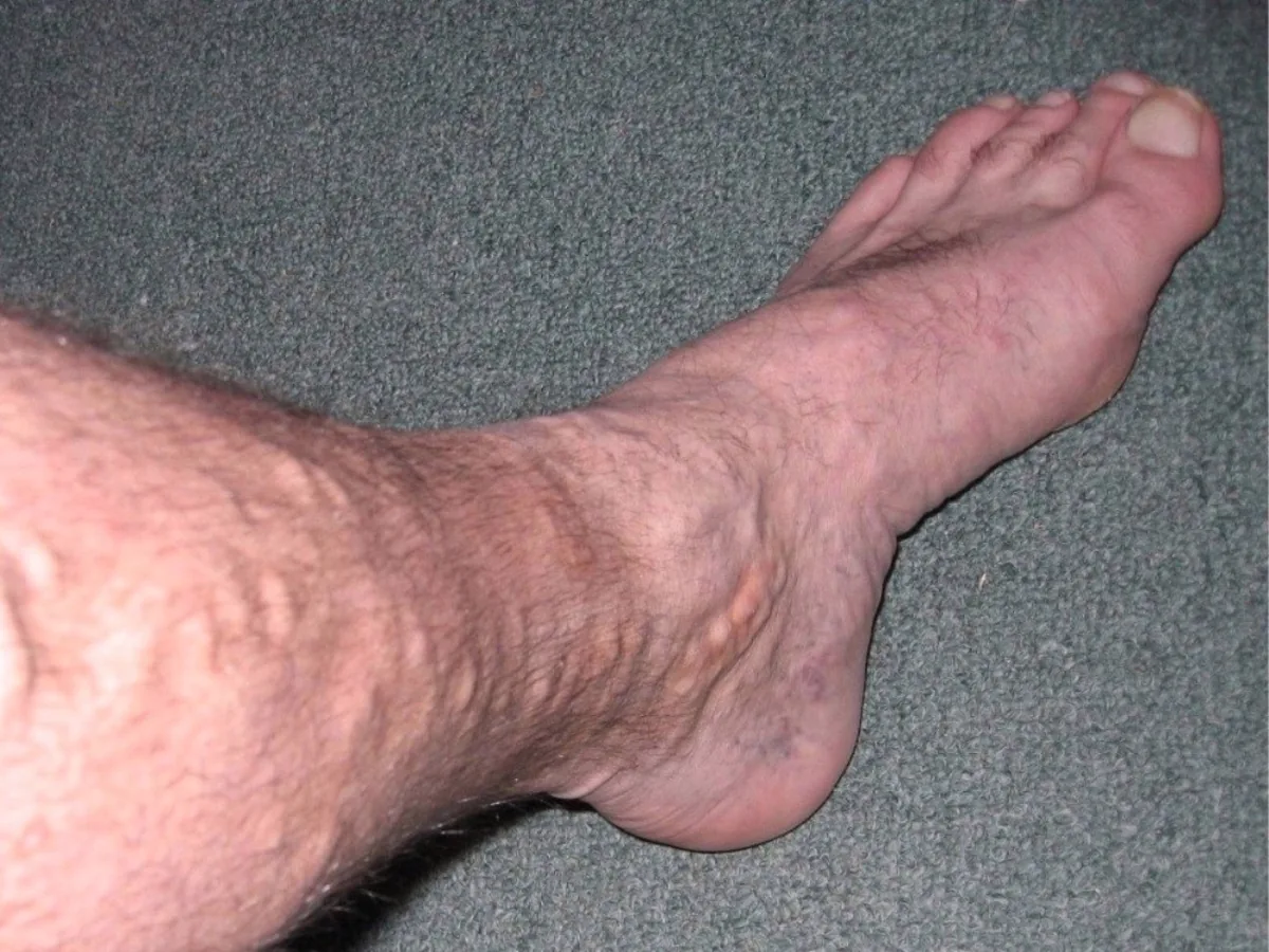
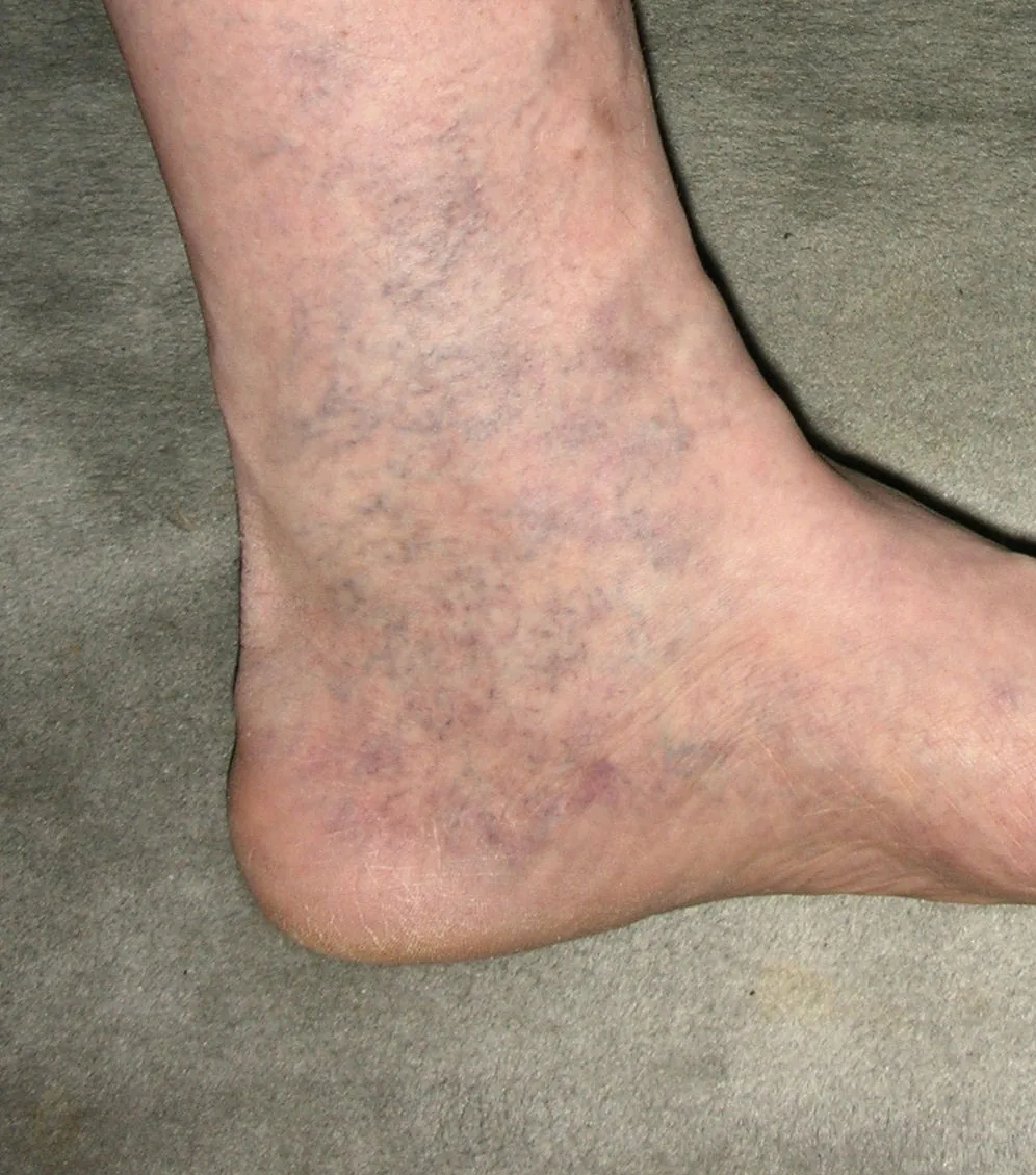
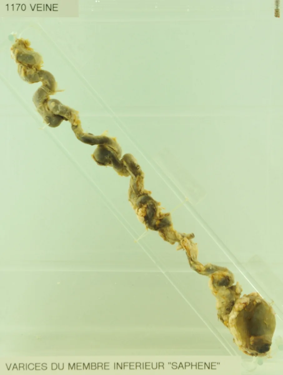 Gross specimen
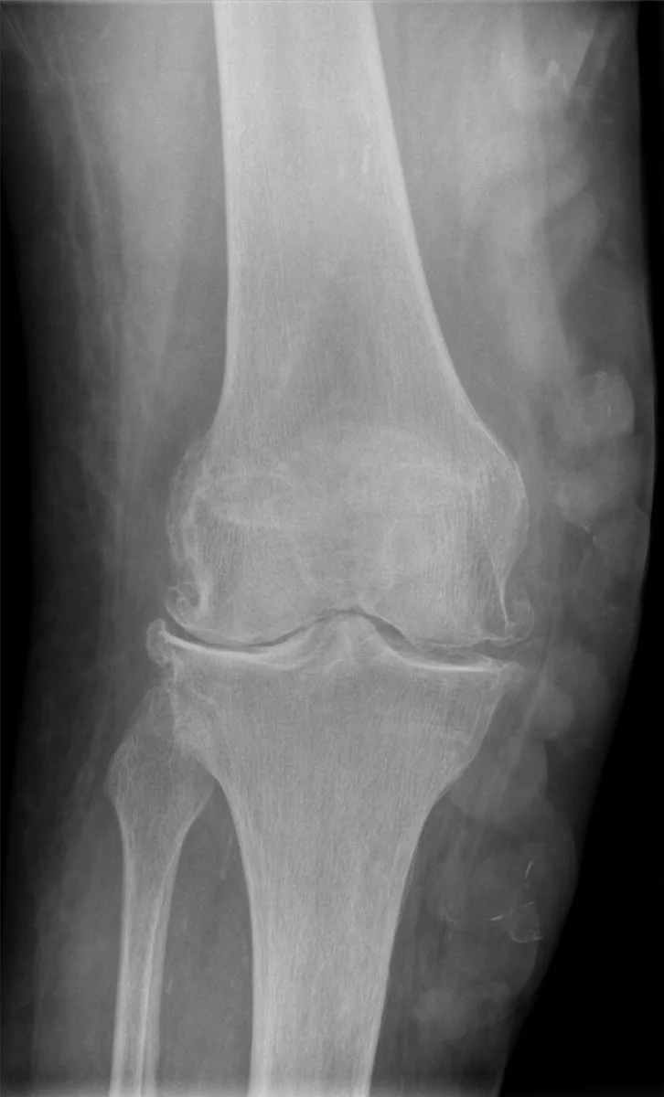 X-ray
X-rayDefinition
Primary varicose veins are a degenerative disease of the vein wall in the superficial venous system of the legs, in which dilated, tortuous veins (varicose veins, varices) develop over the course of life. It is a lifelong, progressive but not life-threatening condition.
They are distinguished from secondary varicose veins, which form as superficial collateral circulations when the deep venous system is obstructed (e.g. after DVT).
Classification
By location and form, the following are distinguished:
- truncal varices of the great or small saphenous vein, including the accessory veins
- tributary varices
- perforator varices
- pelvic varices
- reticular veins
- spider veins (telangiectasias)
In truncal vein incompetence starting at the junction, the Hach classification grades the extent by the length of the reflux segment down to the distal point of incompetence. There are also incomplete truncal varicosis, ascending varicosis without junctional incompetence and special forms (e.g. pudendal, gluteal, pelvic varicosis). Clinically, varicose veins correspond to CEAP class C2; with edema or skin changes, chronic venous insufficiency (C3–C6) is present.
Occurrence & epidemiology
Primary varicose veins are very common. In the Bonn Vein Study, 12.4 % of men and 15.8 % of women had varicose veins without signs of chronic venous insufficiency. The disease can begin in childhood; its frequency increases with age. Women are affected more often than men.
Aetiopathogenesis
A hereditary predisposition is assumed for primary varicose veins; they cluster in families. They result from primary incompetence of the venous valves with reflux or from primary dilatation of the vein wall due to structural weakness.
Main risk factors are:
- older age
- female sex (effect of estrogen on the vein wall, increased venous pressure in the pelvis and legs during pregnancy)
- pregnancies
- positive family history
- orthostatic load (prolonged standing)
The central disease mechanism is disturbed venous hemodynamics with ambulatory venous hypertension. Rarely, varicose veins are part of Klippel-Trénaunay-Weber syndrome with congenital arteriovenous fistulas and capillary angiomas of the skin.
Clinical features
Varicose veins may initially be tense and palpable but not yet visible; later they enlarge, protrude and are most visible when standing. Many cause no symptoms. The most common symptoms are heaviness and fullness, swelling and itching, less often pain after prolonged standing or sitting, and hypersensitivity of the skin.
Complications:
- superficial vein thrombosis (varicophlebitis): a painful, palpable cord
- variceal bleeding: thin-walled superficial venous blebs can rupture and bleed after minimal trauma
- chronic venous insufficiency with edema, trophic skin changes and venous leg ulcers, especially in truncal and perforator varicosis
- increased risk of deep vein thrombosis, particularly in connection with superficial vein thrombosis
Diagnosis
- History: onset and symptoms, swelling, skin changes, itching, previous thromboses, family history
- Clinical examination while standing: visible varices on the thighs and lower legs, edema, hyperpigmentation, eczema, (healed) ulcers; palpable cord-like indurations indicate past or acute superficial vein thrombosis
- Duplex ultrasound: the primary imaging method, performed standing with provocation maneuvers (Valsalva maneuver, squeezing of the calf); it shows reflux in truncal veins and tributaries, the proximal point of incompetence, perforator veins and the state of the deep venous system
- CW Doppler ultrasound: for orientation when duplex ultrasound is unavailable; ankle-brachial index when coexisting peripheral artery disease is suspected
- Functional tests: photoplethysmography (light reflection rheography) and venous occlusion plethysmography
- Phlebography, CT or MR venography: only for special questions (e.g. angiodysplasia, pelvic varicosis)
Keep learning in the app
Further reading (open access)
Cross-references
Note: Learning content for medical education – not a treatment recommendation and no substitute for diagnosis or treatment decisions in individual cases. Treatment and management are deliberately not covered on this page.