Carotid artery stenosis
Board exam relevance: in 1 of 105 exam reports · rank 181- Synonyms
- carotid artery disease, narrowing of the carotid artery, internal carotid artery stenosis, ICA stenosis
- Specialty
- Internal medicine · Angiology
- Images
- Ultrasound 1 · CT 1 · Gross specimen 1
- Last updated
- 10/2026 · Dr. Pascal Bafteh
Contents
Images (3)
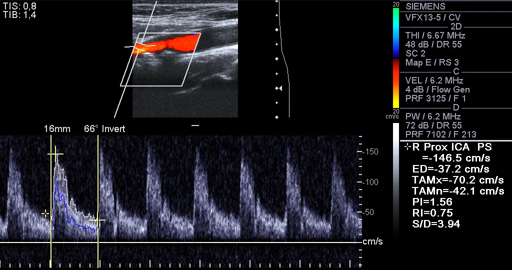 Ultrasound
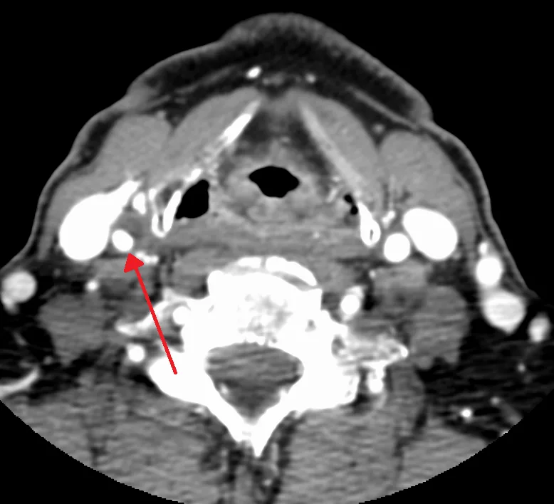 CT
CT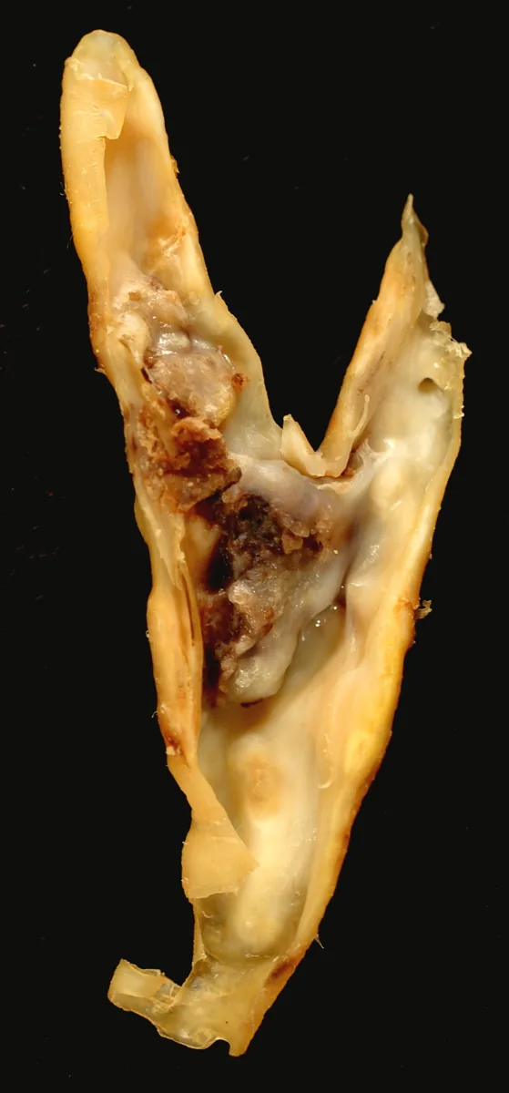 Gross specimen
Definition
Carotid stenosis is a narrowing of the carotid artery supplying the brain, usually the internal carotid artery at the carotid bifurcation, mainly due to atherosclerotic plaques. A stenosis of the internal carotid artery is generally considered significant from a degree of 50 % according to NASCET.
Carotid stenosis is considered symptomatic if it has caused a cerebral infarction, a transient ischemic attack (TIA) or retinal ischemia within the last 6 months; otherwise it is asymptomatic.
Classification
The degree of stenosis is expressed as a reduction in diameter:
- NASCET (distal degree of stenosis): residual lumen compared with the diameter of the internal carotid artery distal to the stenosis; now used almost exclusively
- ECST (local degree of stenosis): residual lumen compared with the original vessel diameter at the level of the stenosis; gives higher values (e.g. 50 % by NASCET corresponds to about 70 % by ECST, 70 % by NASCET to about 80 % by ECST)
Stenoses below 50 %, 50–69 % and 70–99 % as well as occlusion are usually distinguished.
Occurrence & epidemiology
The prevalence of carotid stenosis of at least 50 % in adults is about 4 %; from the age of 65 it rises to 6–15 %. High-grade stenoses (over 70 %) are found in about 1.7 % (pooled). Men are affected more often than women: from the age of 70, the prevalence of stenosis over 50 % in a meta-analysis was 12.5 % in men and 6.9 % in women.
In Germany, about 15 % of all cerebral ischemic events are caused by stenoses of at least 50 % or occlusions of the extracranial carotid artery.
Aetiopathogenesis
The cause is overwhelmingly atherosclerosis. Older age, male sex, known vascular disease (e.g. peripheral artery disease, coronary artery disease, abdominal aortic aneurysm), raised systolic blood pressure, an unfavourable ratio of total to HDL cholesterol, diabetes mellitus and, above all, current smoking are associated with a higher prevalence.
Carotid-related cerebral ischemia is usually caused by emboli of plaque material or thrombi; hemodynamic hypoperfusion beyond a high-grade stenosis is considerably less common. Plaque ulceration and intraplaque hemorrhage can be shown by CT and MRI.
Clinical features
Most carotid stenoses cause no symptoms and are discovered incidentally, for example during ultrasound examinations or because of a bruit.
The spectrum of symptomatic stenosis ranges from retinal circulatory disturbances and transient cerebral ischemia to severe stroke:
- Amaurosis fugax: transient monocular blindness, usually lasting less than 5 minutes, when the ophthalmic artery (a branch of the internal carotid artery) is affected
- TIA: sudden, transient neurological deficits, usually lasting 2–30 minutes, with complete resolution; successive attacks from the carotid territory are usually similar
- Ischemic stroke in the territory of the affected carotid artery, e.g. with hemiparesis or speech disturbance
Diagnosis
- History and neurological examination: the first step in distinguishing symptomatic from asymptomatic stenosis
- Color duplex ultrasound: the primary examination, performed by experienced sonographers; assesses the degree of stenosis and the morphology and extent of the plaque
- DEGUM criteria: a combination of several ultrasound parameters (including peak systolic velocity at the point of maximal stenosis, post-stenotic velocity, collaterals and the ICA/CCA ratio) allows estimation of the degree of stenosis in 10 % steps; the peak systolic velocity is about 200 cm/s at 50 % NASCET and about 300 cm/s at 70 %
- Transcranial Doppler/duplex ultrasound: assessment of hemodynamic compensation
- MR or CT angiography: when the grading is in doubt or ultrasound is difficult
- Imaging of the brain (CT, MRI): detection of infarcts; a fresh, clinically silent ipsilateral ischemic lesion can mark the stenosis as symptomatic
Keep learning in the app
Further reading (open access)
Cross-references
Note: Learning content for medical education – not a treatment recommendation and no substitute for diagnosis or treatment decisions in individual cases. Treatment and management are deliberately not covered on this page.