Lymphedema
- Synonyms
- lymphatic oedema, swollen arm after breast cancer, elephantiasis, Milroy disease
- Specialty
- Internal medicine · Angiology
- Images
- Clinical 3
- Last updated
- 10/2026 · Dr. Pascal Bafteh
Contents
Images (3)
Definition
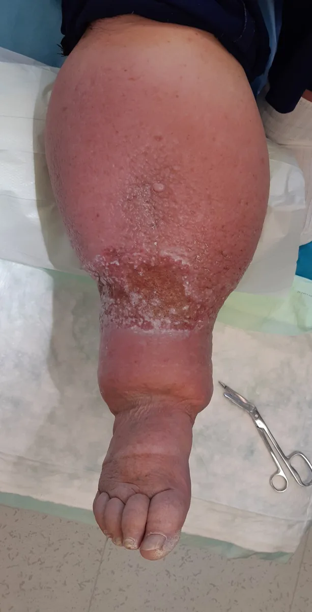
Lymphedema is swelling of a limb (less often of other body regions) due to impaired lymphatic drainage. Primary lymphedema results from maldevelopment (hypoplasia or aplasia) of the lymphatic vessels, secondary lymphedema from obstruction or disruption of previously healthy lymphatic vessels. In the chronic stage the edema is firm, fibrotic and no longer pitting.
Classification
Primary lymphedema is classified by age at onset:
- congenital lymphedema: before the age of 2 years; an autosomal dominant familial form is Milroy disease, usually with bilateral leg edema (mutations in the VEGFR-3 gene)
- lymphedema praecox: between 2 and 35 years, typically in women at the onset of menstruation or in pregnancy (now often called Meige disease)
- lymphedema tarda: after the age of 35
Based on the edema, three stages can be distinguished:
- Stage 1: pitting edema, often resolving overnight
- Stage 2: non-pitting edema; chronic inflammation leads to early fibrosis
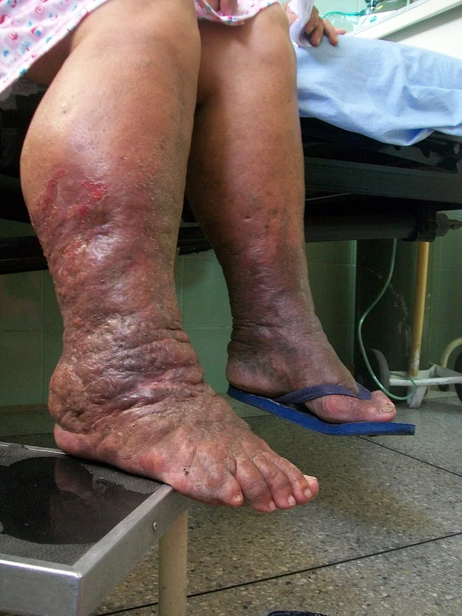
- Stage 3: firm, irreversible edema due to fibrosis; thickened skin, sometimes with an orange-peel appearance
Occurrence & epidemiology
Secondary lymphedema accounts for the vast majority of all lymphedema; primary lymphedema is often inherited, comparatively rare and mostly affects the legs. In the USA lymphadenectomy and radiogenic damage in cancer patients are the most common cause of secondary lymphedema, whereas in tropical and subtropical regions it is lymphatic filariasis.
Aetiopathogenesis
Causes of secondary lymphedema:
- lymphadenectomy or radiogenic damage in the lymphatic drainage area after cancer
- lymphatic filariasis (in tropical and subtropical regions)
- chronic venous insufficiency, in which more lymph leaks into the tissue
- vascular trauma
- obstruction of lymphatic channels by tumors
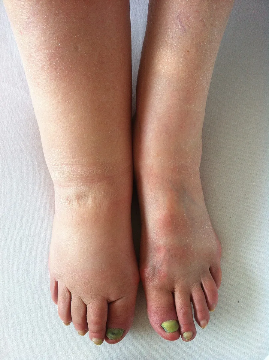
Lymphedema also occurs in some genetic syndromes, such as lymphedema-distichiasis syndrome, yellow nail syndrome (yellow nails, pleural effusions) and Hennekam syndrome.
Clinical features
Symptoms include an aching discomfort and a sensation of heaviness or fullness. The cardinal sign is soft-tissue edema. The swelling is usually unilateral and worsens in warm weather, before menstruation and after prolonged dependency of the limb. It can affect parts of or the entire limb and restrict movement near joints. The physical and emotional burden can be considerable.
Other skin changes include hyperkeratosis, hyperpigmentation, warts, papillomas and fungal infections. Rarely, the limb (or the scrotum) becomes extremely large with severe hyperkeratosis (elephantiasis), more often in filariasis.
Complications:
- Lymphangitis and erysipelas: usually streptococcal, often entering through skin cracks between the toes in athlete's foot; warm, red limb, sometimes with red streaks extending proximally and lymph node swelling
- Cellulitis with skin breakdown
- Lymphangiosarcoma (Stewart-Treves syndrome): very rare in long-standing lymphedema, usually after mastectomy, occasionally in filariasis
Diagnosis
- History and examination: the diagnosis of secondary lymphedema is usually evident from the findings; in primary lymphedema the family history and typical edema are key
- Kaposi-Stemmer sign: a positive sign at the toes indicates a lymphatic component
- Ultrasound: exclusion of other causes of leg swelling, especially deep vein thrombosis
- Lymphoscintigraphy: when the diagnosis or cause is unclear; shows sluggish lymph flow, lymphatic aplasia, abnormal uptake into lymph nodes and signs of lymphatic hypertension
- CT and MRI: secondary tissue changes and sites of lymphatic obstruction; MR lymphography can show lymphatic malformations directly
- Genetic testing: when primary or syndromic lymphedema is suspected
- Testing for filariasis: in people from or staying in endemic areas
- Unexpectedly marked or delayed lymphedema after breast cancer may indicate a tumor recurrence
Keep learning in the app
Further reading (open access)
Cross-references
Note: Learning content for medical education – not a treatment recommendation and no substitute for diagnosis or treatment decisions in individual cases. Treatment and management are deliberately not covered on this page.