Chronic venous insufficiency
Board exam relevance: in 1 of 105 exam reports · rank 181- Synonyms
- CVI, venous insufficiency, post-thrombotic syndrome, venous stasis, chronic venous disease
- Specialty
- Internal medicine · Angiology
- Images
- Clinical 2
- Last updated
- 10/2026 · Dr. Pascal Bafteh
Contents
Images (2)
Definition
Chronic venous insufficiency (CVI) is impaired venous return from the legs with persistent venous hypertension, which can cause symptoms, edema and skin changes. In the CEAP classification it comprises clinical classes C3 to C6.
Post-thrombotic syndrome is symptomatic chronic venous insufficiency following deep vein thrombosis.
Classification
Clinical classification according to CEAP (C classes):
- C0: no visible or palpable signs of venous disease
- C1: telangiectasias and/or reticular veins
- C2: varicose veins
- C3: edema
- C4a: pigmentation, eczema
- C4b: lipodermatosclerosis, atrophie blanche
- C4c: corona phlebectatica (fan-shaped small skin veins at the ankle and foot)
- C5: healed venous ulcer
- C6: active venous ulcer
If symptoms are present, an “s” is added (e.g. C3s). In addition, etiology (E), anatomy (A) and pathophysiology (P) can be described. The CEAP classification is not a severity grading.
Occurrence & epidemiology
In the Bonn Vein Study, one in six men and one in five women had CVI. Venous edema was found in 11.6 % of men and 14.9 % of women, advanced CVI (C4–C6) in 3.8 % of men and 3.4 % of women. Post-thrombotic syndrome frequently develops after deep vein thrombosis, especially after proximal and extensive thrombosis.
Aetiopathogenesis
Venous return from the legs depends on the calf muscle pump and on functioning venous valves. CVI develops when venous obstruction (e.g. after DVT), valvular incompetence, impaired lymphatic flow or reduced muscle pump function (e.g. with immobility) decreases forward flow and increases venous pressure. Fluid overload, for example in right heart failure, also contributes to venous hypertension. Persistently raised pressure leads to edema, inflammation and tissue hypoxia; through incompetent perforator veins it is transmitted to the superficial veins.
Common risk factors:
- deep vein thrombosis
- older age
- obesity
- trauma
- occupations requiring prolonged standing or carrying heavy loads
- pregnancy
Unexplained cases are often attributed to an unnoticed previous DVT. Risk factors for post-thrombotic syndrome are proximal thrombosis, recurrent DVT in the same leg, older age, female sex and estrogen use.
Clinical features
Symptoms include a sense of fullness, heaviness and tension, pain, cramps, tiredness and paraesthesia in the legs. They worsen with standing and walking and improve with rest and elevation; itching may accompany the skin changes.
Findings range from edema to stasis dermatitis on the lower legs and ankles up to ulceration:
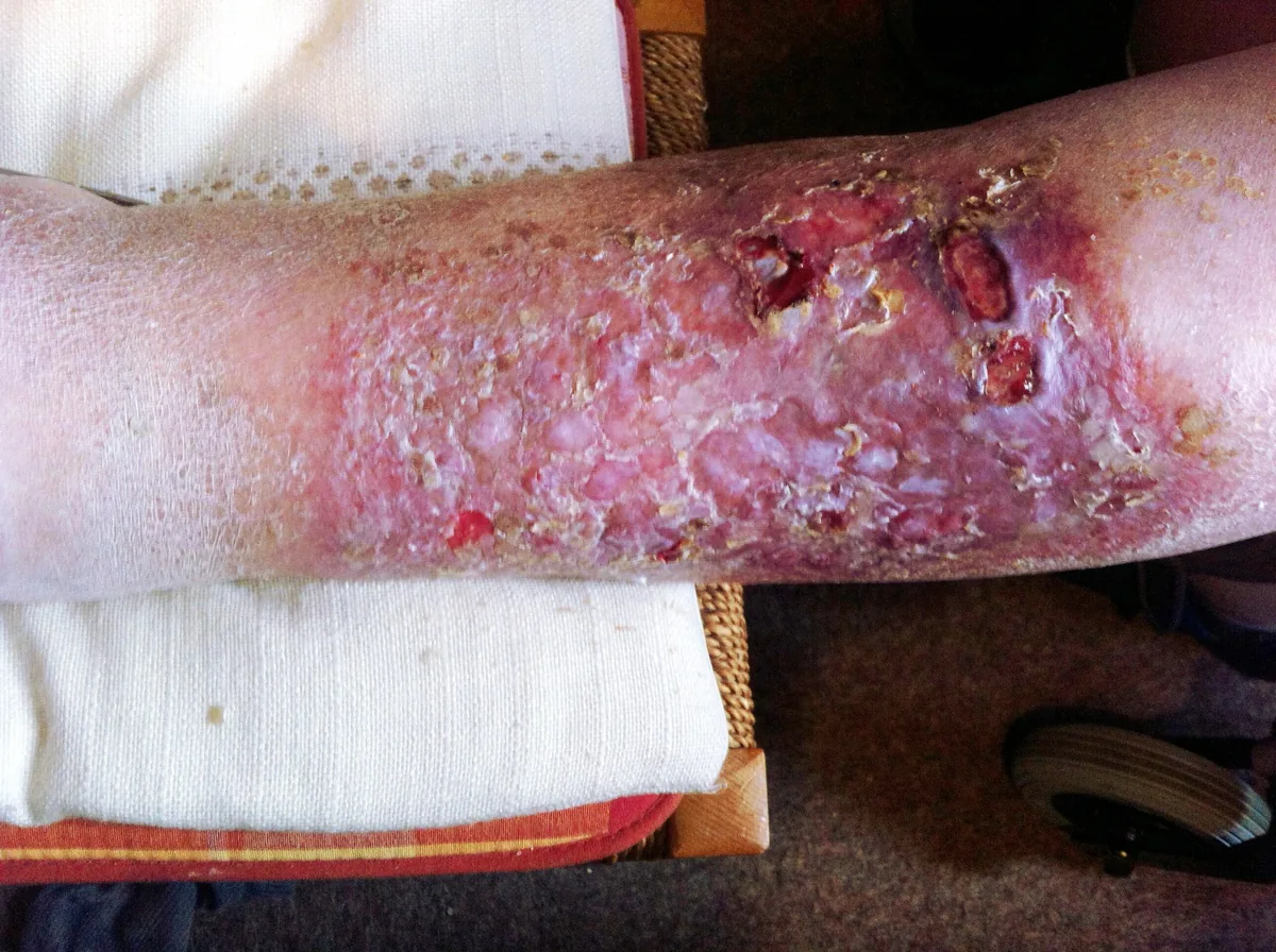
- Stasis dermatitis: erythema, hyperpigmentation, induration, venous ectasia, lichenification
- Lipodermatosclerosis: inflammatory induration with discoloration and edema
- Atrophie blanche: small, white, depressed scars
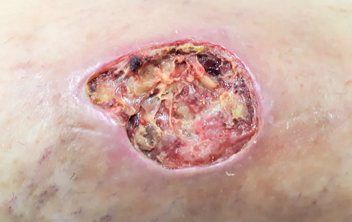
- Venous leg ulcer: typically at the medial malleolus, shallow, moist, sometimes painful; it does not penetrate the deep fascia (unlike arterial ulcers, which may expose tendons or bone)
Clinically evident CVI always shows signs but need not cause symptoms; post-thrombotic syndrome, by contrast, always causes symptoms.
Diagnosis
- History and clinical examination: the basis of diagnosis; in mobile people the examination is performed standing, looking for varicose veins, edema, hyperpigmentation, eczema, induration and (healed) ulcers
- Villalta score: in post-thrombotic syndrome, rates 5 symptoms (pain, cramps, heaviness, pruritus, paraesthesia) and 6 signs (edema, hyperpigmentation, induration, venous ectasia, blanching hyperaemia, calf pain on pressure) each from 0 to 3 points
- Duplex ultrasound: confirmation or exclusion of DVT and depiction of reflux and obstruction
- Functional tests: photoplethysmography (light reflection rheography) and venous occlusion plethysmography quantify venous dysfunction
- Ankle-brachial index: detection of coexisting peripheral artery disease; absence of edema and a reduced ABI suggest an arterial cause instead
Keep learning in the app
Further reading (open access)
Cross-references
Note: Learning content for medical education – not a treatment recommendation and no substitute for diagnosis or treatment decisions in individual cases. Treatment and management are deliberately not covered on this page.