Deep vein thrombosis
Board exam relevance: in 5 of 105 exam reports · rank 69- Synonyms
- DVT, blood clot in the leg, leg vein thrombosis, venous thrombosis, phlebothrombosis
- Specialty
- Internal medicine · Angiology
- Images
- Ultrasound 1 · Histology 1 · Clinical 2
- Last updated
- 10/2026 · Dr. Pascal Bafteh
Contents
Images (4)
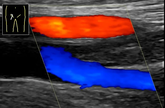 Ultrasound
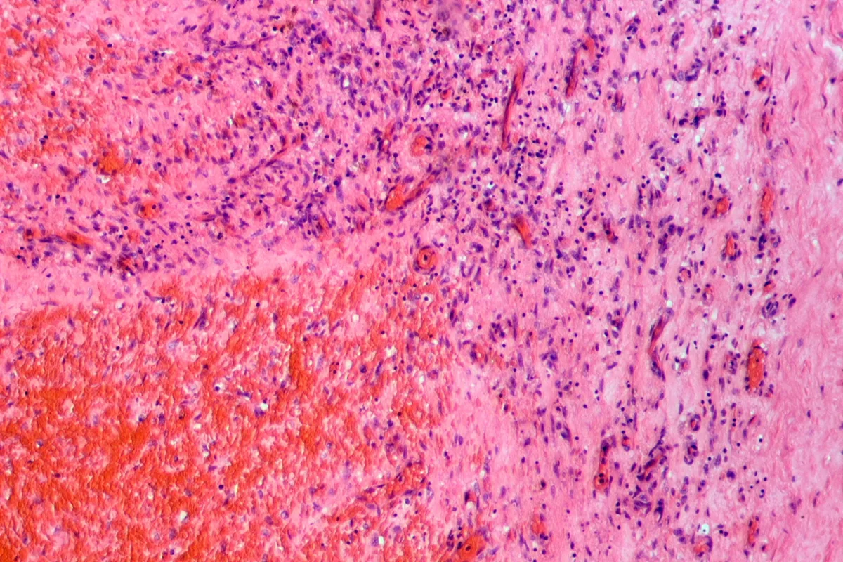 Histology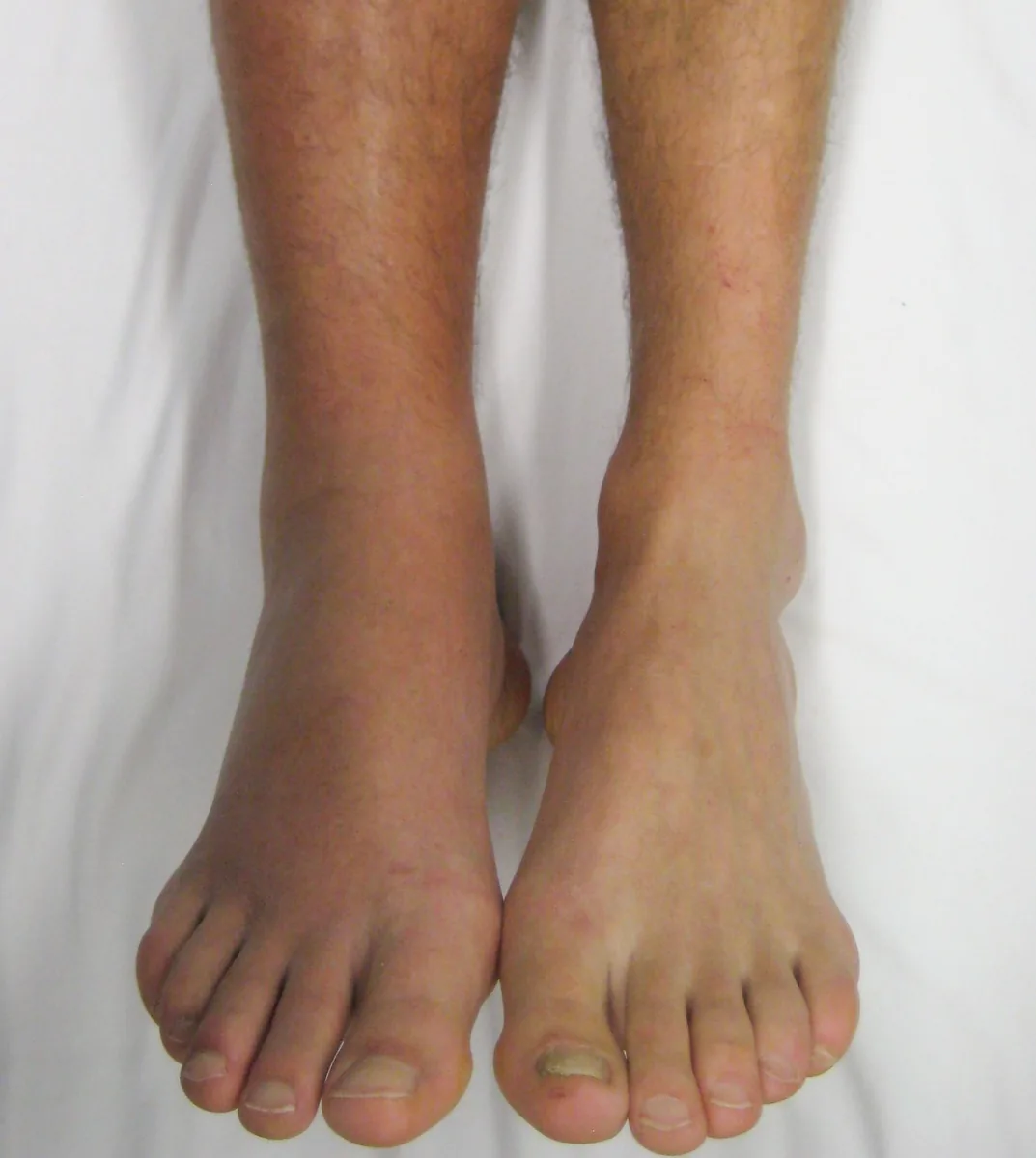
Definition
Deep vein thrombosis (DVT) of the leg and pelvis is a partial or complete occlusion of the deep conducting and/or muscle veins by blood clots (thrombi). It is the main source of pulmonary embolism; both are grouped together as venous thromboembolism (VTE). Thrombosis of the arm veins is much less common (less than 5 % of all DVT).
Classification
- Proximal DVT: involvement of the popliteal vein, femoral vein, common femoral vein, the pelvic veins and/or the inferior vena cava
- Distal DVT: confined to the calf veins; isolated muscle vein thrombosis (gastrocnemius and soleus veins) is distinguished from it
- Ascending thrombosis: the most common course, starting in the calf or muscle veins and growing proximally
- Descending thrombosis: less common, often starting in the pelvic veins, e.g. due to external pressure from a tumor or pregnancy, or in May-Thurner syndrome
- Transfascial thrombosis: spreads from a superficial vein (e.g. the great saphenous vein) to the deep venous system
- Upper-extremity DVT: e.g. caused by central venous catheters or as effort thrombosis (Paget-Schroetter syndrome)
Occurrence & epidemiology
The incidence of DVT is reported as 45–162 per 100,000 person-years. VTE is mainly a disease of older age: the mean age at the first event is about 60 years, and the annual incidence rises from 1–4 per 10,000 at age 20–40 to about 1 per 100 in people over 80. The lifetime prevalence of DVT in adults in Germany is about 3–5 %.
About 40 % of thromboses affect the calf veins, 16 % the popliteal vein, 20 % the femoral veins and 4 % the pelvic veins.
Aetiopathogenesis
The basis is Virchow's triad: impaired venous blood flow (stasis), damage to the vascular endothelium and increased coagulability of the blood. Thrombi usually begin at the venous valves and consist mainly of fibrin and red blood cells (“red thrombus”).
Strong risk factors (odds ratio above 10):
- fractures of the lower limb, major trauma, spinal cord injury
- hip or knee replacement
- hospital stay for heart failure or atrial fibrillation and myocardial infarction within the previous 3 months
- previous venous thromboembolism
Moderate risk factors include cancer (especially with metastases), central venous catheters, oral contraceptives and postmenopausal hormone use, the puerperium, infections, inflammatory bowel disease, autoimmune diseases, stroke with paralysis, superficial vein thrombosis and thrombophilia. Weak risk factors include being bedridden for more than 3 days, long car or air journeys, older age, obesity, pregnancy and varicose veins.
About two thirds of all VTE occur in temporal association with transient risk situations; in up to 30 % no trigger is found. People with cancer have an approximately 7-fold increased risk of VTE, especially with carcinomas of the pancreas, ovary, lung or brain.
Clinical features
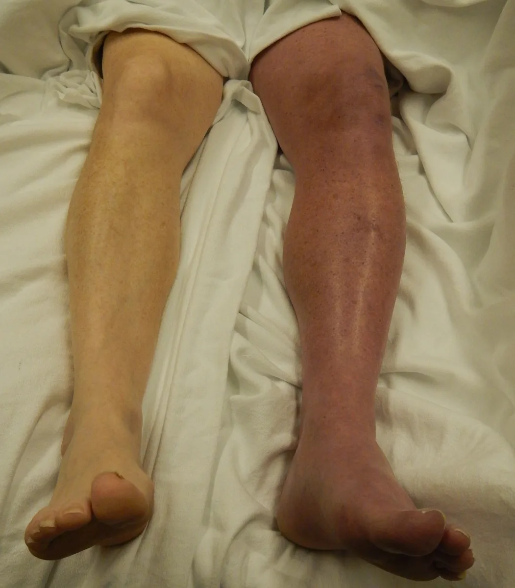
Possible symptoms are leg swelling, pain, a feeling of tension, more prominent superficial veins and cyanosis; low-grade fever may also occur. Dilated superficial collateral veins may become visible.
The classic signs are unreliable:
- Meyer sign: tenderness on the inner side of the lower leg
- Homans sign: calf pain on dorsiflexion of the foot
- Payr sign: pain on pressure on the sole of the foot
In immobilised or severely ill people, thrombosis often causes few or no symptoms.
Complications:
- pulmonary embolism; about half of people with DVT have clinically silent pulmonary embolism
- post-thrombotic syndrome with a tendency to swelling and skin changes up to venous leg ulcers (in about 30 %)
- phlegmasia cerulea dolens: complete blockage of venous outflow with massive, painful, bluish swelling and a risk of compartment syndrome, acute limb ischemia and venous gangrene
- rarely paradoxical embolism through a patent foramen ovale
Diagnosis
Clinical probability
Diagnosis begins with an estimate of the clinical probability, for example with the Wells score (1 point each):
- active cancer
- immobilisation of a leg by a cast spanning a joint or due to paresis
- bedridden for at least 3 days or major surgery within the last 3 months
- tenderness along the deep veins
- swelling of the entire leg
- calf swelling with a circumference at least 3 cm larger than the other side
- unilateral edema in the symptomatic leg
- prominent, non-varicose superficial collateral veins
- previous deep vein thrombosis
- alternative diagnosis at least as likely: minus 2 points
In the two-level assessment, 2 or more points indicate a high and 0–1 points a low clinical probability.
D-dimers
D-dimers are formed during the breakdown of cross-linked fibrin. Normal D-dimers make acute thrombosis unlikely; with a low clinical probability, DVT is thereby considered excluded. Raised values are non-specific and are regularly found after major surgery or trauma, in infections and inflammation, disseminated intravascular coagulation, cancer and pregnancy; they also rise with age.
The usual cut-off is 500 µg/l. From the age of 50, an age-adjusted cut-off (age × 10 µg/l) can be used.
Imaging
- Compression ultrasound: the primary imaging method. The decisive criterion is absent or incomplete compressibility of the vein under pressure from the transducer. A freshly thrombosed vein is often distended and filled with hypoechoic material
- Extent of the examination: either complete with duplex support from the common femoral vein down to the calf veins, or limited to selected points (e.g. groin and popliteal fossa)
- Duplex ultrasound: flow profile in the common femoral vein compared with the other side; loss of respiratory variation in flow points to an obstruction further proximally
- CT or MR venography: if ultrasound is negative or inconclusive and suspicion persists, for example when pelvic vein thrombosis is suspected
- Further work-up: history, physical examination and basic laboratory tests to identify risk factors
Keep learning in the app
Further reading (open access)
Cross-references
Note: Learning content for medical education – not a treatment recommendation and no substitute for diagnosis or treatment decisions in individual cases. Treatment and management are deliberately not covered on this page.