Acute mesenteric ischemia
Board exam relevance: in 3 of 105 exam reports · rank 111- Synonyms
- bowel infarction, mesenteric infarction, intestinal ischaemia, mesenteric artery occlusion, NOMI
- Specialty
- Internal medicine · Angiology
- Images
- CT 2 · Histology 1
- Last updated
- 10/2026 · Dr. Pascal Bafteh
Contents
Images (3)
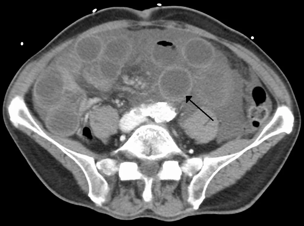 CT
CT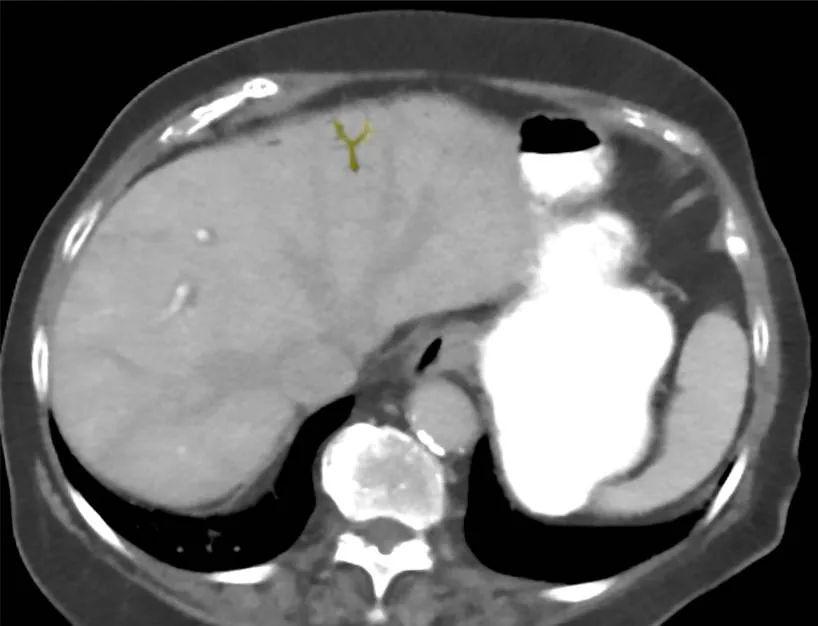 CT
CT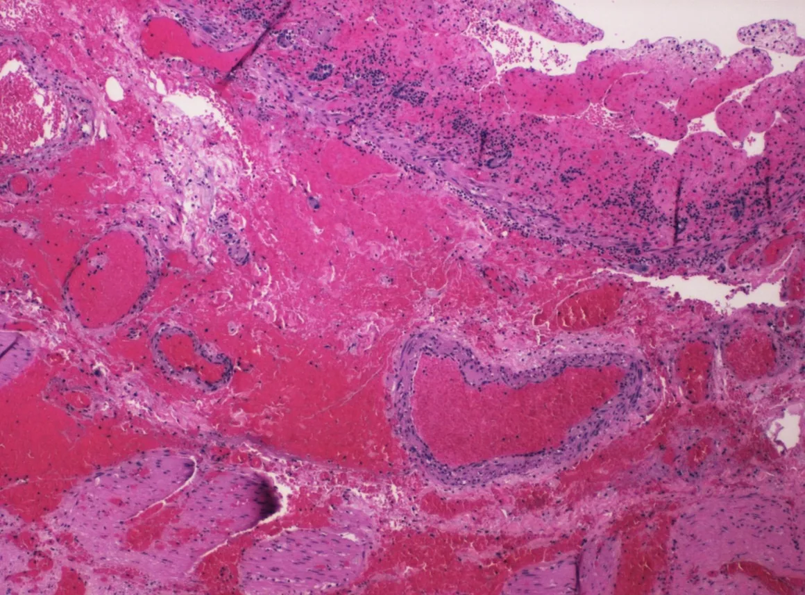 Histology
Definition
Acute mesenteric ischemia is a sudden interruption of intestinal blood flow due to embolism, thrombosis or a low-flow state. It leads to mediator release, inflammation and ultimately intestinal infarction.
It is distinguished from ischemic colitis, which involves only small vessels and mainly causes mucosal necrosis and bleeding.
Classification
By the cause of the occlusion, the following are distinguished:
- arterial embolism (over 40 %)
- arterial thrombosis (about 30 %)
- mesenteric venous thrombosis (about 15 %)
- non-occlusive mesenteric ischemia (NOMI) (about 15 %)
Occurrence & epidemiology
People over 50 years of age with relevant risk factors are at greatest risk; however, many patients have no identifiable risk factors.
Aetiopathogenesis
Three large vessels supply the abdominal organs: the celiac trunk (esophagus, stomach, proximal duodenum, liver, gallbladder, pancreas, spleen), the superior mesenteric artery (distal duodenum, jejunum, ileum and colon up to the splenic flexure) and the inferior mesenteric artery (descending colon, sigmoid, rectum). The stomach, duodenum and rectum have abundant collaterals and rarely become ischemic; the splenic flexure is a watershed between the superior and inferior mesenteric arteries and is particularly at risk.
Risk factors by type of occlusion:
- embolism: coronary artery disease, heart failure, valvular heart disease, atrial fibrillation, previous arterial emboli
- arterial thrombosis: generalised atherosclerosis
- venous thrombosis: hypercoagulable states, inflammatory conditions (e.g. pancreatitis, diverticulitis), trauma, heart failure, renal failure, portal hypertension, decompression sickness
- NOMI: low-flow states (heart failure, shock, heart-lung machine), splanchnic vasoconstriction (e.g. due to vasoactive drugs or cocaine)
The intestinal mucosa has a high metabolic rate and normally receives 20–25 % of cardiac output; it is therefore very sensitive to reduced perfusion. Ischemia destroys the mucosal barrier, releasing bacteria, toxins and vasoactive mediators. Consequences are myocardial depression, a systemic inflammatory response and multiple organ failure. Necrosis can occur as early as 6 hours after the onset of symptoms.
Clinical features
The early hallmark is severe abdominal pain with minimal physical findings: the abdomen remains soft with little or no tenderness; mild tachycardia may be present.
- sudden onset of pain suggests arterial embolism but is not diagnostic
- gradual onset is typical of venous thrombosis
- a history of postprandial abdominal pain (intestinal angina) points to arterial thrombosis
As necrosis progresses, signs of peritonitis develop with marked tenderness, guarding, rigidity and absent bowel sounds. The stool may contain occult blood, increasingly so as ischemia progresses. Finally, signs of shock appear.
Diagnosis
Clinical judgement is more important than individual tests. Mesenteric ischemia is to be considered in anyone over 50 years with risk factors and sudden severe abdominal pain.
- CT angiography or selective mesenteric angiography: the primary investigations; CT can show the vascular occlusion directly, more accurately on the venous side
- MR angiography: when iodinated contrast medium cannot be used; very accurate for proximal, less so for distal occlusions
- Plain abdominal X-ray: mainly to rule out other causes (e.g. perforation); late findings are portal venous gas or gas in the bowel wall (pneumatosis intestinalis)
- Doppler ultrasound: can show arterial occlusion but has low sensitivity
- Laboratory tests: creatine kinase and lactate rise only with necrosis and are non-specific; in the early phase imaging and laboratory values are often still normal
Keep learning in the app
Further reading (open access)
Cross-references
Note: Learning content for medical education – not a treatment recommendation and no substitute for diagnosis or treatment decisions in individual cases. Treatment and management are deliberately not covered on this page.