Pulmonary embolism
Board exam relevance: in 17 of 105 exam reports · rank 11- Synonyms
- PE, blood clot in the lung, pulmonary thromboembolism, lung embolism
- Specialty
- Internal medicine · Angiology
- Images
- CT 2 · ECG 1 · X-ray 1 · Scintigraphy 1 · Gross specimen 1
- Last updated
- 10/2026 · Dr. Pascal Bafteh
Contents
Images (6)
 CT
CT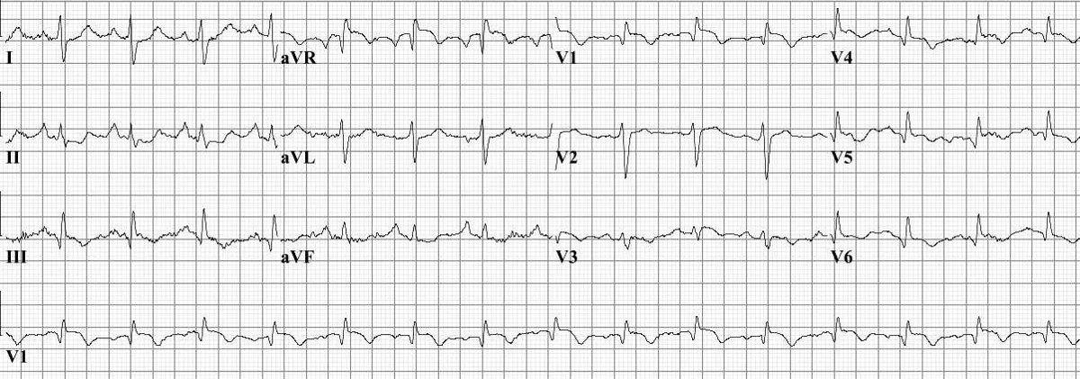 ECG
ECG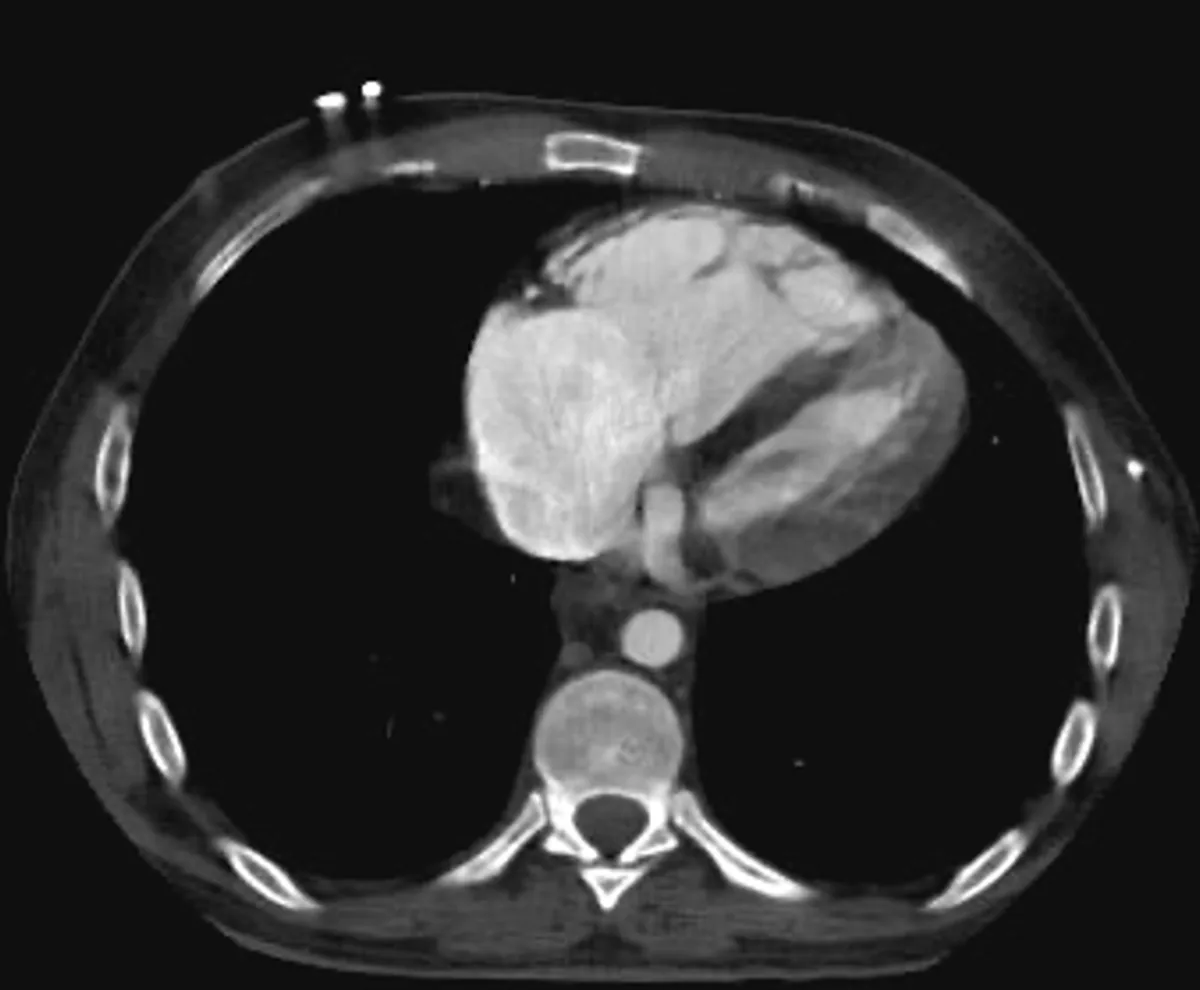 CT
CT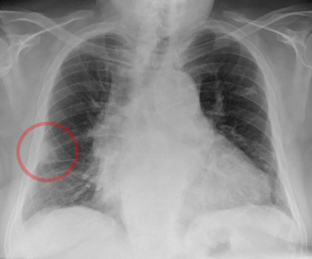 X-ray
X-ray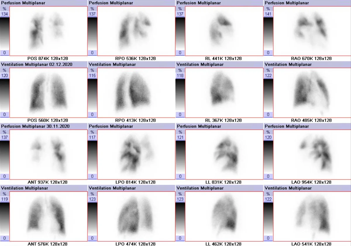 Scintigraphy
 Gross specimen
Definition
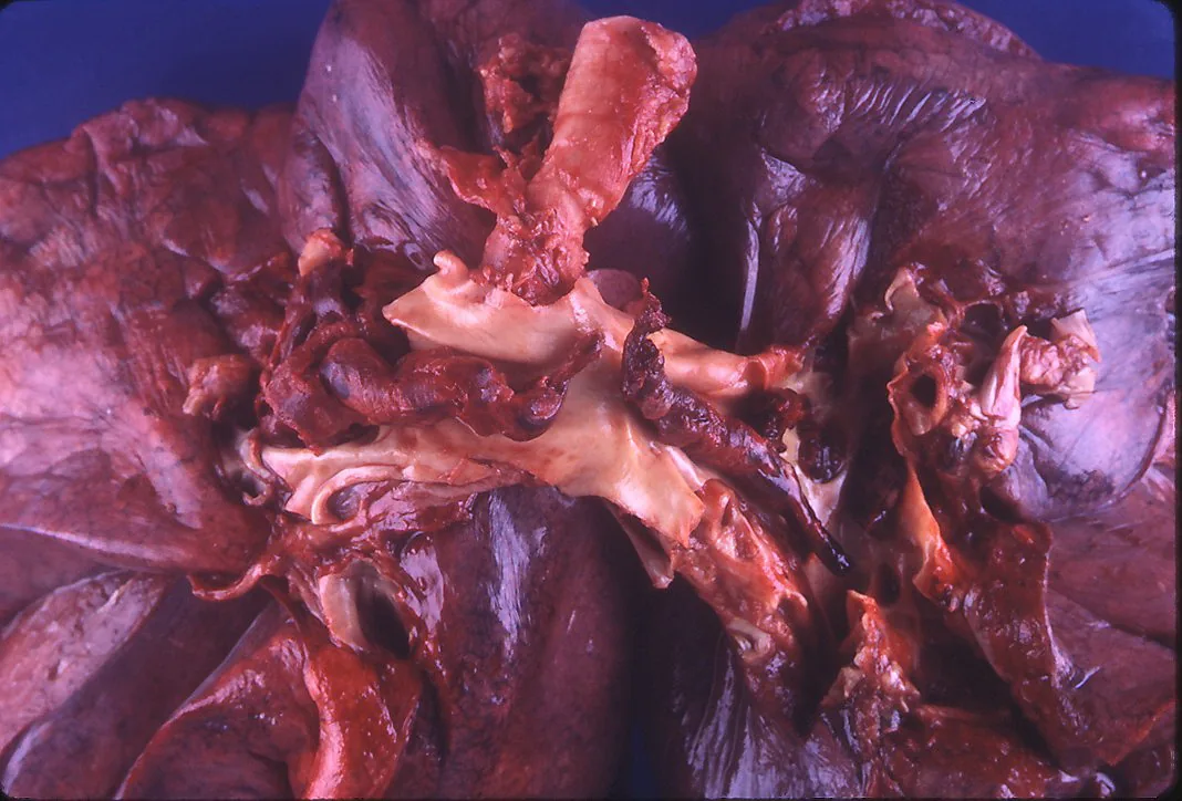
Pulmonary embolism (PE) is a partial or complete occlusion of the pulmonary arteries, usually by thrombi carried from the deep veins of the legs or pelvis. Together with deep vein thrombosis it forms the condition known as venous thromboembolism (VTE). Non-thrombotic emboli (e.g. from air, amniotic fluid, fat or tumor cells) are rare.
Classification
Basic forms
- By hemodynamics: patients with cardiorespiratory arrest, shock or persistent hypotension are considered unstable, all others stable
- By location: central, segmental and subsegmental emboli; a saddle embolus lodges at the bifurcation of the main pulmonary artery
Severity classes (ESC)
In stable patients, severity is further classified according to the ESC. The basis is the simplified Pulmonary Embolism Severity Index (sPESI) with 1 point each for age over 80 years, active cancer, chronic heart or lung disease, heart rate ≥ 110/min, systolic blood pressure below 100 mmHg and arterial oxygen saturation below 90 %.
- high risk: hemodynamically unstable
- intermediate-high: sPESI ≥ 1 with right heart strain on echocardiography or CT and raised cardiac biomarkers
- intermediate-low: sPESI ≥ 1 without this combination
- low: sPESI 0 without signs of right heart strain
Occurrence & epidemiology
In epidemiological studies, the annual incidence is up to 115 per 100,000 people and rises with age. After myocardial infarction and stroke, pulmonary embolism is the third most common acute cardiovascular disease. In women, VTE manifests as pulmonary embolism more often than in men.
Aetiopathogenesis
The risk factors are those of deep vein thrombosis (Virchow's triad: stasis, endothelial injury, hypercoagulability). Thrombi that reach the popliteal vein or extend further proximally embolise more often.
Mechanical obstruction and vasoconstriction raise pulmonary vascular resistance. Right ventricular afterload increases; the ventricle dilates, and tricuspid regurgitation and increased wall stress develop, up to acute right heart failure (acute cor pulmonale). Gas exchange is impaired, resulting in hypoxemia. Many emboli are small and cause no symptoms. Endogenous fibrinolysis reduces most emboli; the physiological changes regress within hours to days.
Pulmonary infarction is mainly caused by smaller, peripherally lodged emboli; because of the dual blood supply of the lung, it is relatively rare.
Clinical features
Symptoms and findings
The symptoms are non-specific. The most common are sudden-onset breathlessness, chest pain (often worse on breathing), syncope or presyncope and hemoptysis. The spectrum ranges from no symptoms to shock and cardiac arrest.
Findings include tachypnea and tachycardia, and distended neck veins in right heart failure. Lung auscultation is usually normal; signs of leg vein thrombosis are often absent.
Complications
- pulmonary infarction, pleural effusion
- acute right heart failure, obstructive shock, sudden cardiac death
- rarely paradoxical embolism through a patent foramen ovale
- chronic thromboembolic pulmonary hypertension (CTEPH) after 1–3 % of pulmonary emboli
Diagnosis
Clinical probability
In stable patients, diagnosis begins with the clinical probability, for example using the Wells score (original points):
- clinical signs of DVT: 3
- PE more likely than an alternative diagnosis: 3
- recent postoperative state or immobilisation: 1.5
- heart rate above 100/min: 1.5
- previous VTE: 1.5
- hemoptysis: 1
- active cancer: 1
A value above 4 indicates a high, 0–4 a low clinical probability. The revised Geneva score is an alternative.
Laboratory tests
- D-dimers: normal D-dimers make pulmonary embolism unlikely when the clinical probability is not high; raised values are non-specific. Above the age of 50, an age-adjusted cut-off (age × 10 µg/l) applies
- Blood gas analysis: hypoxemia, hypocapnia or an increased alveolar–arterial oxygen difference, but often normal
- Troponin, BNP and NT-proBNP: may be raised in right ventricular strain
ECG
Possible findings are sinus tachycardia, T-wave inversion in V1–V4, an S1Q3T3 pattern, right bundle branch block and atrial fibrillation. The S1Q3T3 pattern and a new right bundle branch block occur in only about 5 %. A normal ECG does not rule out pulmonary embolism.
Imaging
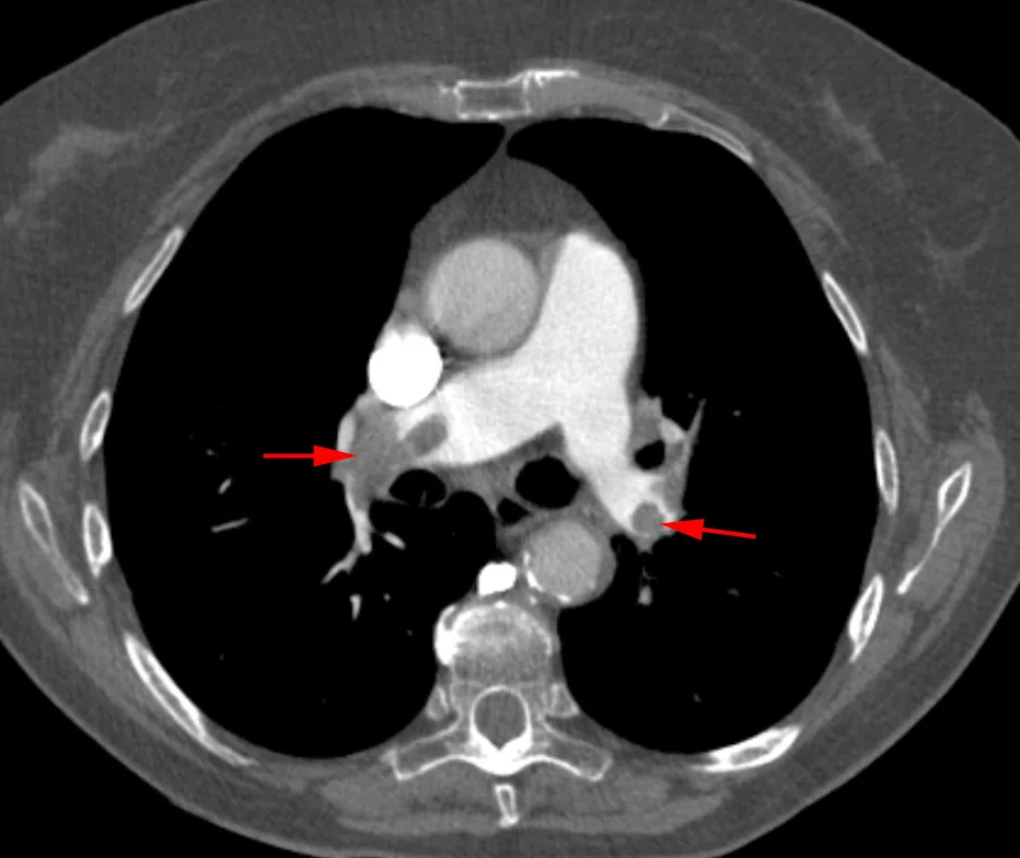
- CT pulmonary angiography (CTPA): the standard method for confirming the diagnosis (sensitivity 94 %, specificity 98 %); a right-to-left ventricular ratio above 1.0 indicates right heart strain
- Lung scintigraphy (V/Q SPECT): a fallback method in stable patients (sensitivity 92 %, specificity 91 %); areas with preserved aeration but reduced perfusion (mismatch) are typical
- Chest X-ray: usually non-specific
- Compression ultrasound of the leg veins: detection of DVT supports the diagnosis
Echocardiography
Echocardiography shows signs of right heart strain: a dilated right ventricle (ratio to the left ventricle above 1), a flattened interventricular septum (D sign), reduced TAPSE (normal ≥ 17 mm) and the McConnell sign (free-wall hypokinesia with preserved apical function). In unstable patients it is the key bedside examination: if signs of right heart strain are absent, massive pulmonary embolism is an unlikely cause. A normal echocardiogram does not rule out a smaller embolism.
Keep learning in the app
Further reading (open access)
Cross-references
Note: Learning content for medical education – not a treatment recommendation and no substitute for diagnosis or treatment decisions in individual cases. Treatment and management are deliberately not covered on this page.