Aortic dissection
Board exam relevance: in 3 of 105 exam reports · rank 111- Synonyms
- aortic tear, dissecting aneurysm, acute aortic syndrome, tear in the aorta
- Specialty
- Internal medicine · Angiology
- Images
- CT 2 · X-ray 1
- Last updated
- 10/2026 · Dr. Pascal Bafteh
Contents
Images (3)
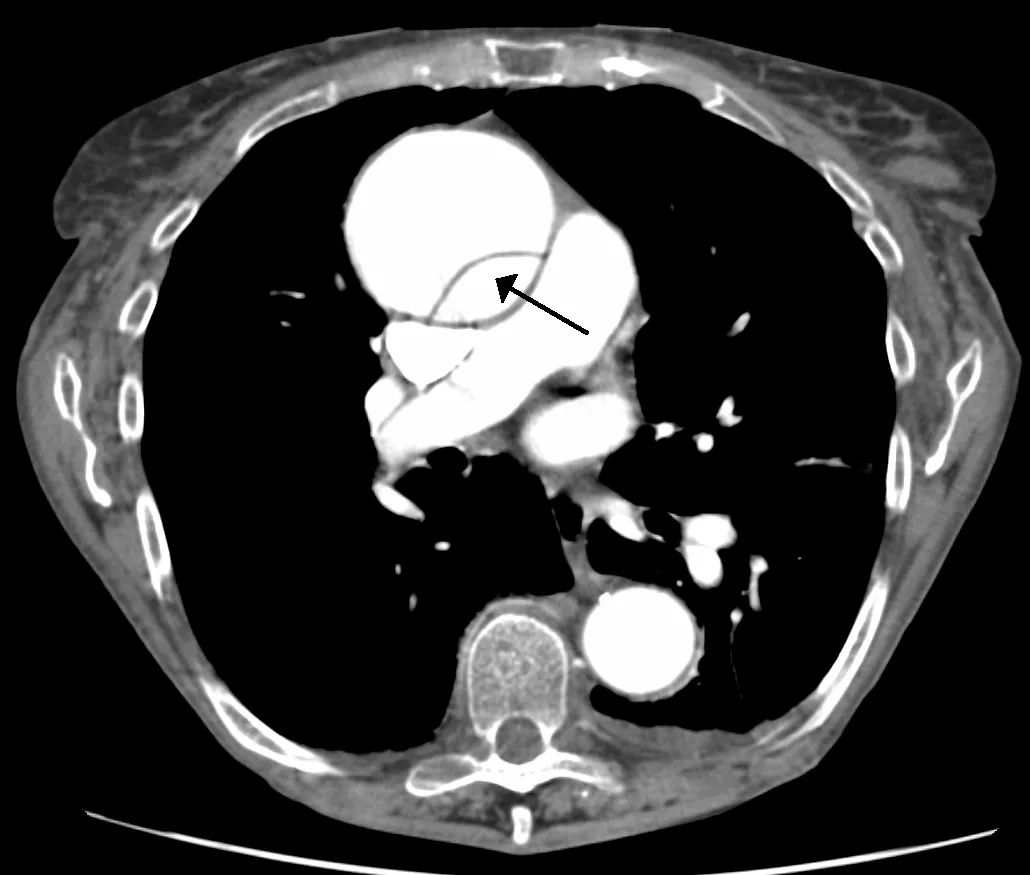 CT
CT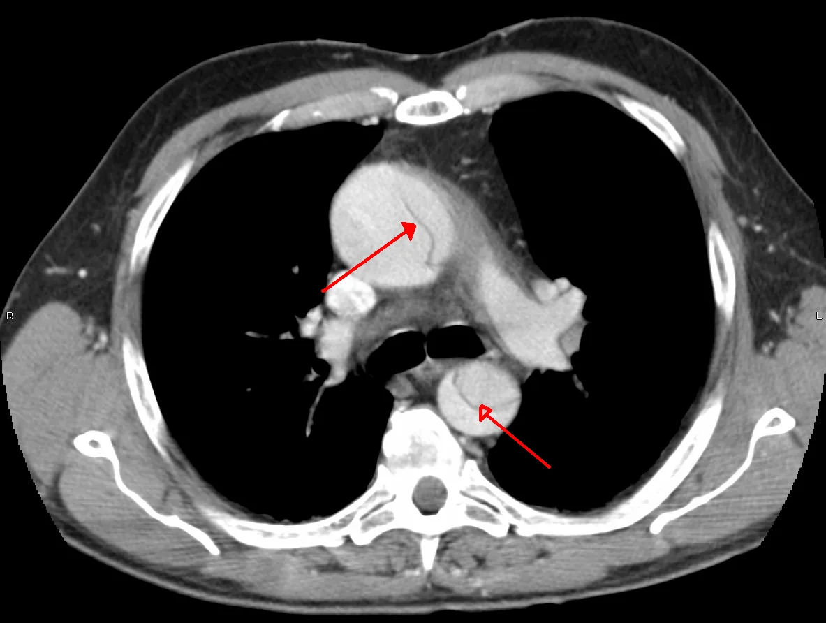 CT
CT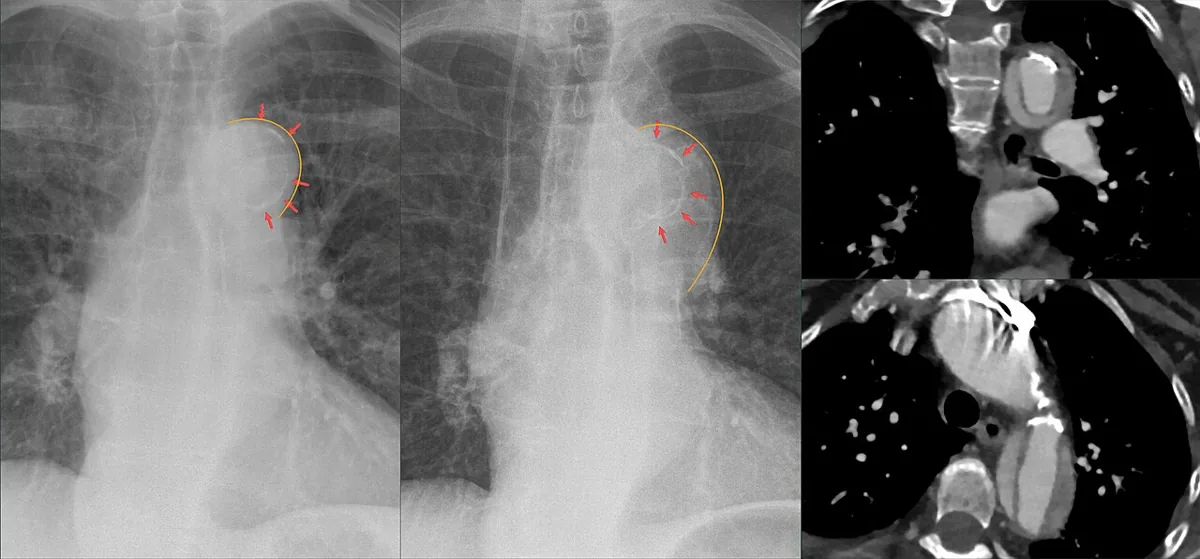 X-ray
X-rayDefinition
In aortic dissection, blood flows through a tear in the intima into the aortic wall, separates the intima from the media and creates a false lumen. The intimal tear may be a primary event or secondary to hemorrhage within the media. The dissection can arise anywhere along the aorta and extend proximally or distally into branch arteries.
Variants or precursors are intramural hematoma without a visible intimal tear, an intimal tear with bulging but no false lumen, and dissection or hematoma caused by an ulcerating atherosclerotic plaque.
Classification
Stanford classification:
- Type A: involvement of the ascending aorta
- Type B: confined to the descending aorta (distal to the origin of the left subclavian artery)
DeBakey classification:
- Type I: starts in the ascending aorta and extends at least to the aortic arch, often beyond (most common type)
- Type II: starts in and is confined to the ascending aorta
- Type III: starts in the descending aorta distal to the left subclavian artery; IIIa is confined to the thoracic aorta, IIIb extends below the diaphragm
In terms of timing, dissections present for less than two weeks are considered acute; they most often cause complications.
Occurrence & epidemiology
Men, older people, people with arterial hypertension and Black people are at particular risk. Peak incidence in the general population is at 50–65 years, and at 20–40 years in congenital connective tissue disorders (e.g. Marfan or Ehlers-Danlos syndrome).
Aetiopathogenesis
Pre-existing degeneration of the aortic media is often present. Causes and contributing factors are:
- atherosclerotic risk factors (involved in more than two thirds): above all arterial hypertension, as well as smoking, dyslipidemia and cocaine use
- congenital or hereditary connective tissue disorders: bicuspid aortic valve, coarctation of the aorta, cystic medial necrosis, Ehlers-Danlos, Loeys-Dietz, Marfan and Turner syndrome, familial thoracic aortic aneurysm
- iatrogenic: e.g. catheterisation of the aorta
- systemic inflammatory diseases: Behçet disease, giant cell arteritis, Takayasu arteritis
- trauma: deceleration injuries
Pathophysiologically, inflammation of the aortic wall, loss of smooth muscle cells, degeneration of the media and disruption of elastic fibers lead to dissection. It most often starts at sites of greatest hydraulic stress: the right lateral wall of the ascending aorta (within 5 cm of the aortic valve) and the proximal descending aorta just beyond the origin of the left subclavian artery.
Clinical features
Typical is a sudden, excruciating pain behind the sternum or between the scapulae, often described as tearing. The pain frequently migrates as the dissection progresses. Up to 20 % of patients have syncope; hypotension and tachycardia may indicate active bleeding.
Further findings:
- Pulse deficits: in about 20–30 %, sometimes waxing and waning; blood pressure differences between the limbs, sometimes over 30 mmHg
- Aortic regurgitation: often a diastolic murmur in proximal dissection, rarely acute heart failure
- Cardiac tamponade: pulsus paradoxus and distended neck veins
- Left pleural effusion due to leaking blood or exudate
- Malperfusion: stroke, myocardial infarction (typically inferior infarction when the right coronary artery is involved), intestinal infarction, renal insufficiency with oliguria or anuria, paraparesis or paraplegia, limb ischemia
- Rupture of the aorta into the pericardium, right atrium or left pleural space
Diagnosis
Aortic dissection is to be considered in chest or back pain, unexplained syncope, unexplained abdominal pain, stroke or acute heart failure, especially with unequal pulses or blood pressures in the limbs.
- Chest X-ray: often a widened mediastinum, frequently with a localised bulge at the site of origin; left pleural effusion is common
- CT angiography: usually the first imaging method (sensitivity 90–100 %, specificity 87–100 %); an intimal flap and a double lumen confirm the diagnosis
- Transesophageal echocardiography: sensitivity 97–99 %, possible at the bedside in less than 20 minutes and without contrast agents
- MR angiography: nearly 100 % sensitive and specific, but time-consuming; suited to stable patients with subacute or chronic symptoms
- Echocardiography: detection of aortic regurgitation
- ECG: not diagnostic of the dissection itself; signs of inferior infarction together with a new aortic regurgitation murmur suggest a type I dissection involving the right coronary artery
- Laboratory tests: troponin and CK-MB help to distinguish it from myocardial infarction; mild leukocytosis and anemia with blood loss; raised LDH as a non-specific sign of celiac or mesenteric artery involvement
Keep learning in the app
Further reading (open access)
Cross-references
Note: Learning content for medical education – not a treatment recommendation and no substitute for diagnosis or treatment decisions in individual cases. Treatment and management are deliberately not covered on this page.