Abdominal aortic aneurysm
Board exam relevance: in 4 of 105 exam reports · rank 90- Synonyms
- AAA, aortic aneurysm, bulge in the abdominal aorta, triple A
- Specialty
- Internal medicine · Angiology
- Images
- CT 2 · Angiography 1
- Last updated
- 10/2026 · Dr. Pascal Bafteh
Contents
Images (3)
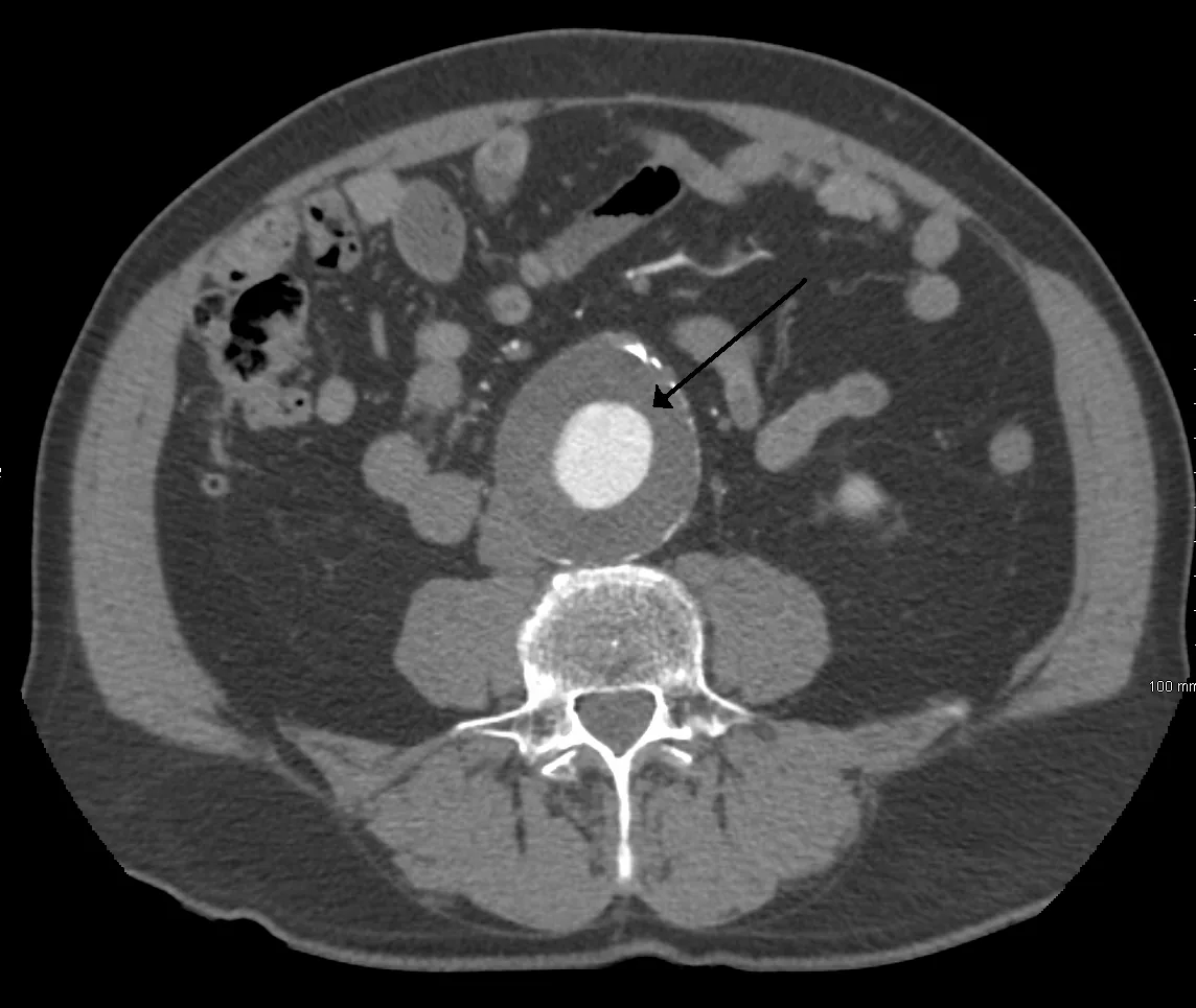 CT
CT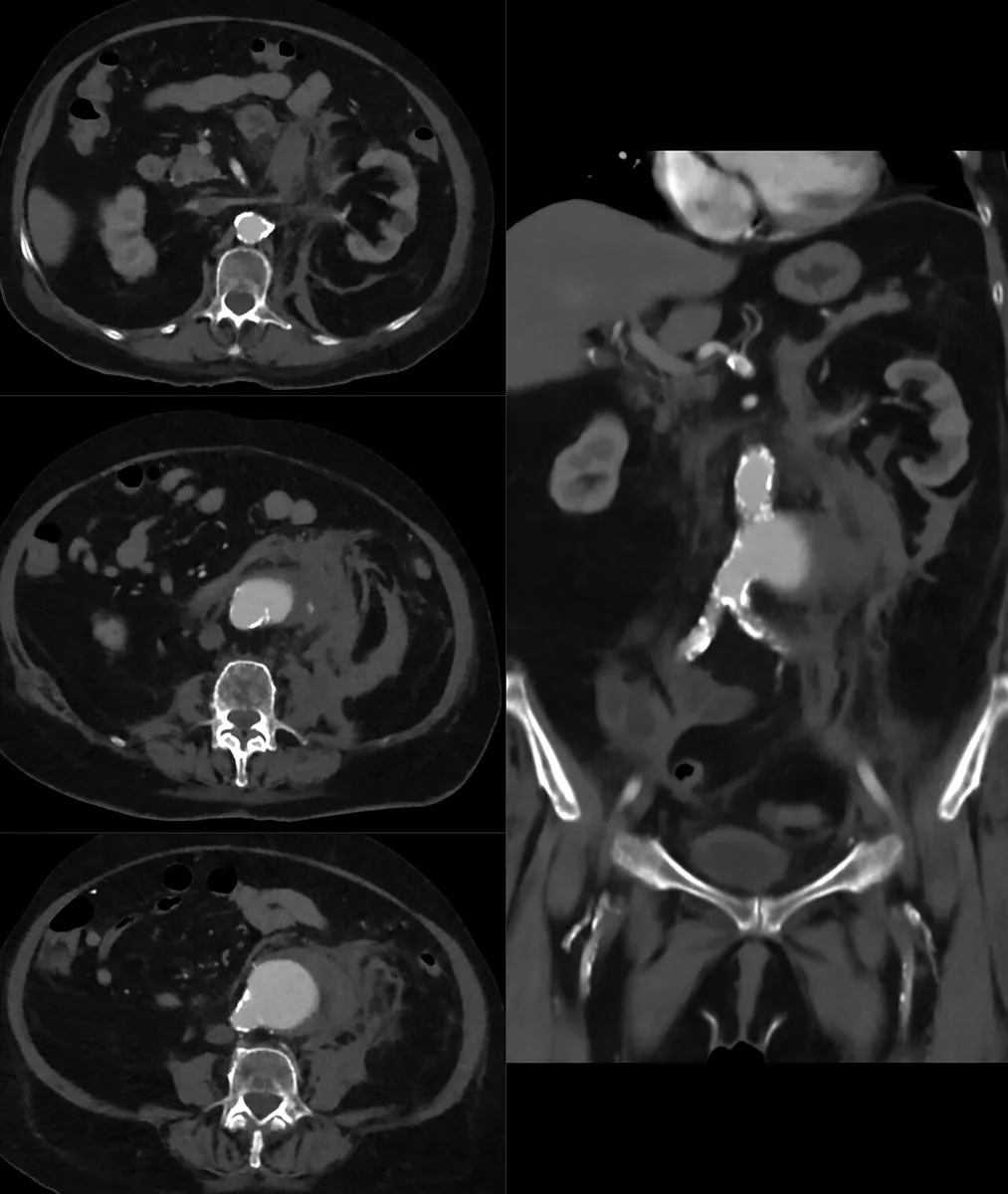 CT
CT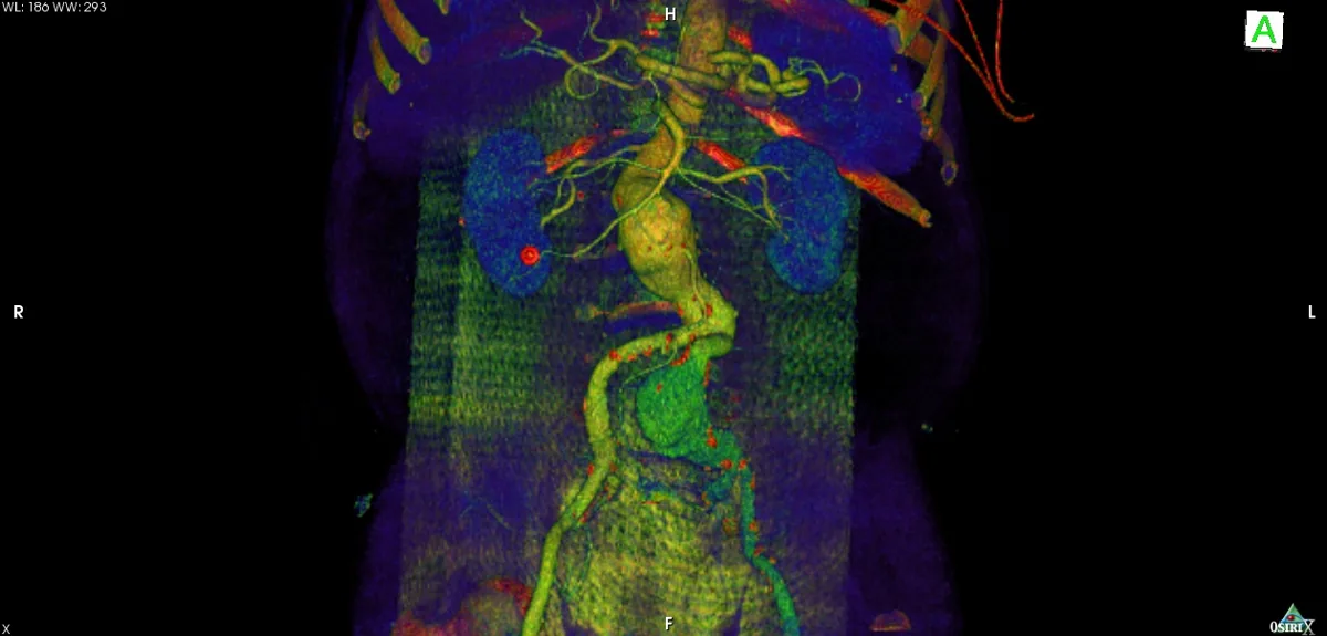 Angiography
Definition
An abdominal aortic aneurysm (AAA) is defined as an abdominal aortic diameter of 3 cm or more. Abdominal aortic aneurysms account for about three quarters of all aortic aneurysms. They typically begin below the renal arteries (infrarenal) but may include the renal artery origins (pararenal); about half extend into the iliac arteries.
Classification
- By shape: usually spindle-shaped (fusiform, circumferential widening), less often saccular; many are lined with laminated thrombus
- By location: infrarenal, pararenal (including the renal artery origins), with involvement of the iliac arteries
- By cause: degenerative-atherosclerotic, inflammatory (vasculitis), infected (“mycotic”), traumatic, in connective tissue disorders
- By clinical status: asymptomatic, symptomatic, ruptured
Occurrence & epidemiology
Depending on the population studied, 0.4–7.6 % are affected. Men are about three times more likely to be affected than women. The disease occurs mainly at the age of 70 to 80 years and is more common in White people than in people of African ancestry.
Aetiopathogenesis
The cause is multifactorial; usually the aortic wall is weakened, most commonly by atherosclerosis. Other causes include:
- trauma
- cystic medial necrosis (degeneration of the aortic media)
- anastomotic disruption of vascular grafts
- vasculitides (e.g. Takayasu arteritis)
- rarely syphilis or bacterial or fungal infection in sepsis or endocarditis (mycotic aneurysm; most common organizm Staphylococcus aureus, followed by Salmonella)
Risk factors:
- smoking (the strongest risk factor)
- arterial hypertension
- older age
- male sex
- positive family history (in 15–25 %)
- inherited connective tissue disorders (e.g. Marfan, Ehlers-Danlos, Loeys-Dietz syndrome)
Growth is usually slow (about 10 % per year), often stepwise with periods of no growth; the risk of rupture rises with size.
Clinical features
Most abdominal aortic aneurysms cause no symptoms and are discovered incidentally. Symptoms usually result from pressure on adjacent structures:
- steady, deep, boring pain, mainly in the lumbosacral region
- an abnormally prominent abdominal pulsation
- a rapidly enlarging aneurysm that is about to rupture may be tender
On examination, the aneurysm may be palpable as a pulsatile mass, depending on its size and body habitus; a systolic bruit may be audible over it.
Complications:
- Rupture: most often on the left posterolateral wall 2–4 cm below the renal arteries; typical are abdominal or back pain, hypotension and tachycardia, often after minor trauma or straining (e.g. lifting)
- Embolisation: dislodged thrombus or atheromatous material can occlude renal, intestinal and leg arteries
- Disseminated intravascular coagulation (uncommon)
Diagnosis
- Clinical examination: a palpable pulsatile mass is of limited value; only when its diameter exceeds 5 cm does the probability that an aneurysm is actually present exceed 80 %
- Abdominal ultrasound: detection and measurement of size; in unstable patients with suspected rupture it is the fastest bedside test, but limited by bowel gas
- CT or MRI of the abdomen: confirmation of the diagnosis
- CT or MR angiography: precise depiction of size and anatomy; with mural thrombus, conventional angiography may underestimate the true size, whereas CT estimates it more accurately
- Plain abdominal X-ray: neither sensitive nor specific, but may show calcification of the aneurysm wall
- Blood cultures: when a mycotic aneurysm is suspected
An abdominal aortic aneurysm is to be considered in older people with acute abdominal or back pain, even if no pulsatile mass is palpable.
Keep learning in the app
Further reading (open access)
Cross-references
Note: Learning content for medical education – not a treatment recommendation and no substitute for diagnosis or treatment decisions in individual cases. Treatment and management are deliberately not covered on this page.