Raynaud's phenomenon
Board exam relevance: in 2 of 105 exam reports · rank 142- Synonyms
- Raynaud's disease, Raynaud syndrome, white finger, cold fingers, vasospasm of the fingers
- Specialty
- Internal medicine · Angiology
- Images
- Clinical 3
- Last updated
- 10/2026 · Dr. Pascal Bafteh
Contents
Images (3)
Definition
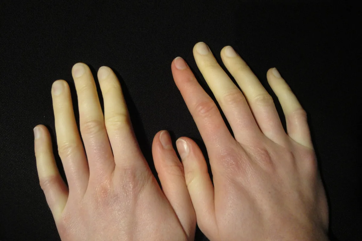
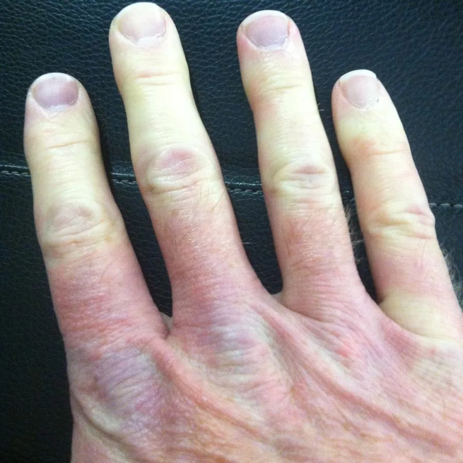
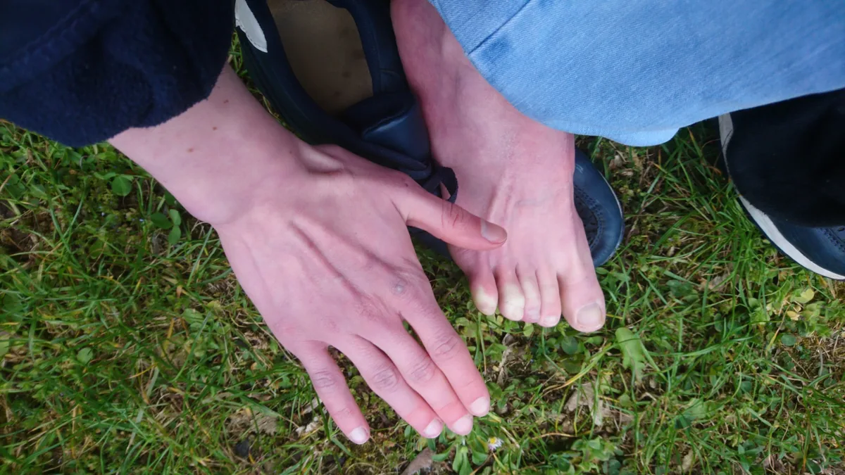
Raynaud's phenomenon is an episodic, reversible spasm of blood vessels (vasospasm) in the fingers, less often in the toes or other acral parts (e.g. nose, tongue), triggered by cold or emotional stress. It causes discomfort and color changes of the affected digits.
Primary Raynaud's phenomenon (Raynaud's disease) without an underlying condition is distinguished from secondary Raynaud's phenomenon (Raynaud's syndrome) accompanying another disease.
Classification
Features distinguishing primary from secondary forms:
- Onset: primary usually in adolescence and young adulthood; secondary more often in childhood or older age (onset after the age of 40 suggests a secondary form)
- Attacks: primary mild and symmetrical on both hands; secondary often severe, painful, sometimes asymmetrical or unilateral
- Tissue damage: primary no necrosis or ulcers; secondary frequently digital ulcers
- Arterial occlusion: primary no, secondary frequently
- Nailfold capillaroscopy: primary normal or non-specific; secondary, depending on the underlying disease, e.g. a scleroderma pattern
- Autoantibodies: primary negative or low-titre non-specific; secondary positive with detection of specific antigens
Occurrence & epidemiology
Overall prevalence is about 3–5 %; 5.8 % has been calculated for Germany. Women are affected more often than men, and younger people more often than older people. Raynaud's phenomenon is more common at northern latitudes and in mountainous regions. Primary Raynaud's phenomenon accounts for 80–90 % of cases.
Aetiopathogenesis
An exaggerated α2-adrenergic vascular reaction is thought to trigger the vasospasm. The phenomenon mainly affects skin areas with many arteriovenous anastomoses; local cold on the hands and fingers appears to be decisive.
Causes of secondary Raynaud's phenomenon:
- connective tissue and inflammatory rheumatic diseases: systemic sclerosis, systemic lupus erythematosus, mixed connective tissue disease, polymyositis/dermatomyositis, rheumatoid arthritis, Sjögren syndrome
- endocrine: hypothyroidism
- hematological: cold agglutinin disease, polycythemia vera
- paraneoplastic
- neurological and vascular: carpal tunnel syndrome, thoracic outlet syndrome
- substances: nicotine (often overlooked), cocaine, ergot alkaloids, sympathomimetics
In systemic sclerosis, the vasospasm affects vessels already narrowed by intimal fibrosis. Raynaud's phenomenon may occur together with migraine, variant angina and pulmonary hypertension.
Clinical features
Cold, emotional stress or vibration trigger a sensation of cold, burning pain, paraesthesia and color changes in one or more fingers. All symptoms resolve once the stimulus is removed; rewarming accelerates recovery.
- The color changes are sharply demarcated and often symmetrical
- triphasic: pallor (white), then cyanosis (blue), and after rewarming redness due to reactive hyperaemia (red); also biphasic (blue, red) or uniphasic (only white or blue)
- typically the middle three fingers are affected, rarely the thumb; not proximal to the metacarpophalangeal joints
- duration from minutes to hours
Primary Raynaud's phenomenon rarely causes tissue loss. Secondary Raynaud's phenomenon in connective tissue diseases can lead to painful digital gangrene; in systemic sclerosis, very painful, infected fingertip ulcers often develop.
Diagnosis
The diagnosis is clinical: unusual cold sensitivity and at least biphasic color change of the fingers (white and blue). Unlike Raynaud's phenomenon, acrocyanosis is persistent, not quickly reversible and causes no trophic changes, ulcers or pain.
- History and examination: looking specifically for signs of an underlying disease; pulse status and Allen test to assess the radial and ulnar arteries
- Vascular testing: demonstration of the tendency to vasospasm (digital pulse waveforms and pressures, thermal provocation), color duplex ultrasound to exclude arterial occlusion
- Nailfold capillaroscopy: an important clue to a secondary form, e.g. a scleroderma pattern
- Laboratory tests: ESR or CRP, antinuclear antibodies (ANA) and extractable nuclear antigens (ENA), anti-DNA, anticentromere and anti-Scl-70 antibodies, rheumatoid factor and anti-CCP antibodies
- Search for cancer: with B symptoms or first occurrence in older age
Keep learning in the app
Further reading (open access)
Cross-references
Note: Learning content for medical education – not a treatment recommendation and no substitute for diagnosis or treatment decisions in individual cases. Treatment and management are deliberately not covered on this page.