Microscopic colitis
Board exam relevance: in 1 of 105 exam reports · rank 181- Synonyms
- collagenous colitis, lymphocytic colitis, watery diarrhoea, chronic diarrhoea
- Specialty
- Internal medicine · Gastroenterology
- Images
- Histology 3
- Last updated
- 10/2026 · Dr. Pascal Bafteh
Contents
Images (3)
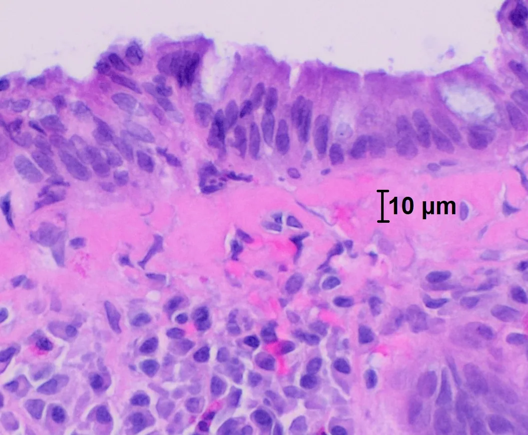 Histology
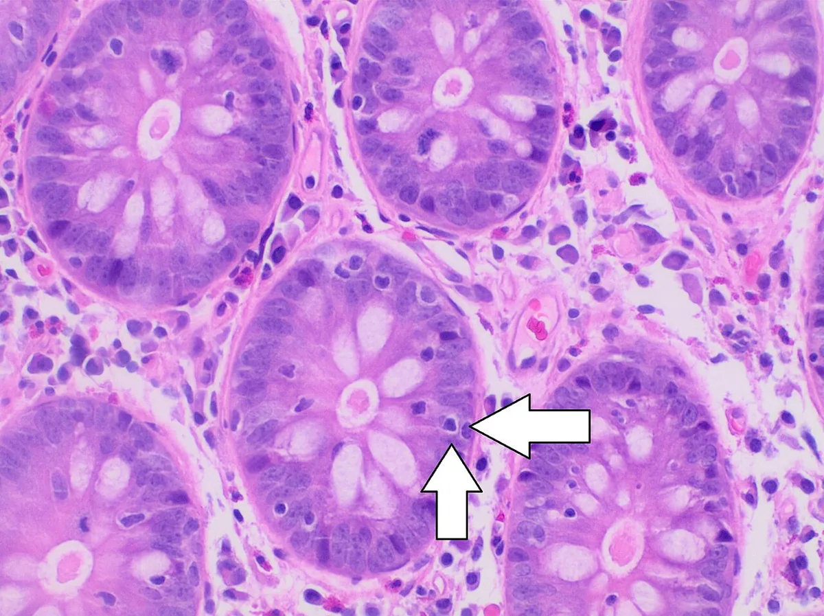 Histology
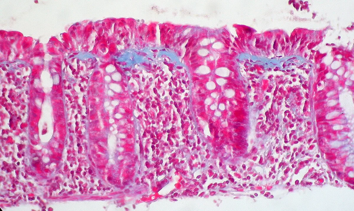 Histology
Definition
Microscopic colitis is an inflammatory bowel disease, probably caused by a chronic immune-mediated process, and a common cause of chronic watery non-bloody diarrhea. It is characterised by the triad of chronic watery diarrhea, normal mucosal appearance at colonoscopy and typical histology. Collagenous and lymphocytic colitis are distinguished.
Classification
Histological criteria (European guideline 2021, hematoxylin and eosin staining):
- Collagenous colitis: thickened subepithelial collagen band of more than 10 µm.
- Lymphocytic colitis: at least 20 intraepithelial lymphocytes per 100 surface epithelial cells with an increased inflammatory infiltrate of the lamina propria, without a significantly thickened collagen band (< 10 µm).
- Incomplete collagenous colitis: collagen band over 5 µm but under 10 µm.
- Incomplete lymphocytic colitis: more than 10 but fewer than 20 intraepithelial lymphocytes with a normal collagen band.
Occurrence & epidemiology
Incidence is estimated at about 11.4 per 100,000 person-years and is increasing. The mean age at diagnosis is 60–65 years; women are affected much more often, with a female-to-male ratio of up to 9:1.
Aetiopathogenesis
The cause is unknown and probably multifactorial. An abnormal immune reaction to luminal antigens in genetically predisposed individuals is proposed.
- Genetic predisposition.
- Altered gut microbiota with reduced diversity.
- Immunological processes: e.g. an increase in CD8-positive lymphocytes in the epithelium and lamina propria.
- Bile acid malabsorption: bile acids increase chloride secretion, reduce fluid absorption and increase colonic motility.
- Autoimmune diseases: frequently coexisting, e.g. celiac disease.
- Smoking and certain drugs: acid-suppressing drugs in particular are associated with the disease.
Clinical features
- Chronic, watery, non-bloody diarrhea as the typical cardinal symptom.
- Pattern of involvement: all segments of the colon except the rectum may be affected; involvement is uneven and patchy and usually most severe in the proximal colon.
- Laboratory findings: about half of patients have mild anemia.
Histology
Both forms show a mild inflammatory infiltrate of the lamina propria. Collagenous colitis is characterised by the thickened subepithelial collagen band, lymphocytic colitis by the increase in intraepithelial lymphocytes. Because the changes are patchy and most marked proximally, rectal biopsies have the highest rate of false-negative results.
Diagnosis
- History: travel, recent intake of antibacterial drugs (risk of Clostridioides difficile), drugs and dietary changes.
- Stool tests: Clostridioides difficile and stool cultures to exclude infection; fecal calprotectin or lactoferrin to distinguish other inflammatory bowel diseases.
- Laboratory tests: no laboratory test establishes the diagnosis; blood count and electrolytes are not helpful for diagnosis or screening.
- Colonoscopy with biopsies: the diagnosis rests on histology. Usually at least eight biopsies (two or more each from the right, transverse, left and sigmoid colon) or at least six biopsies (three each from the ascending and descending colon) are taken. If no biopsies are taken above the rectosigmoid colon, about one in five cases of microscopic colitis is missed; samples from sigmoidoscopy alone have low sensitivity.
Keep learning in the app
Further reading (open access)
Cross-references
More topics: Gastroenterology
Note: Learning content for medical education – not a treatment recommendation and no substitute for diagnosis or treatment decisions in individual cases. Treatment and management are deliberately not covered on this page.