Achalasia
Board exam relevance: in 3 of 105 exam reports · rank 111- Synonyms
- oesophageal achalasia, esophageal achalasia, cardiospasm, megaoesophagus, swallowing disorder
- Specialty
- Internal medicine · Gastroenterology
- Images
- X-ray 2 · CT 1
- Last updated
- 10/2026 · Dr. Pascal Bafteh
Contents
Images (3)
 X-ray
X-ray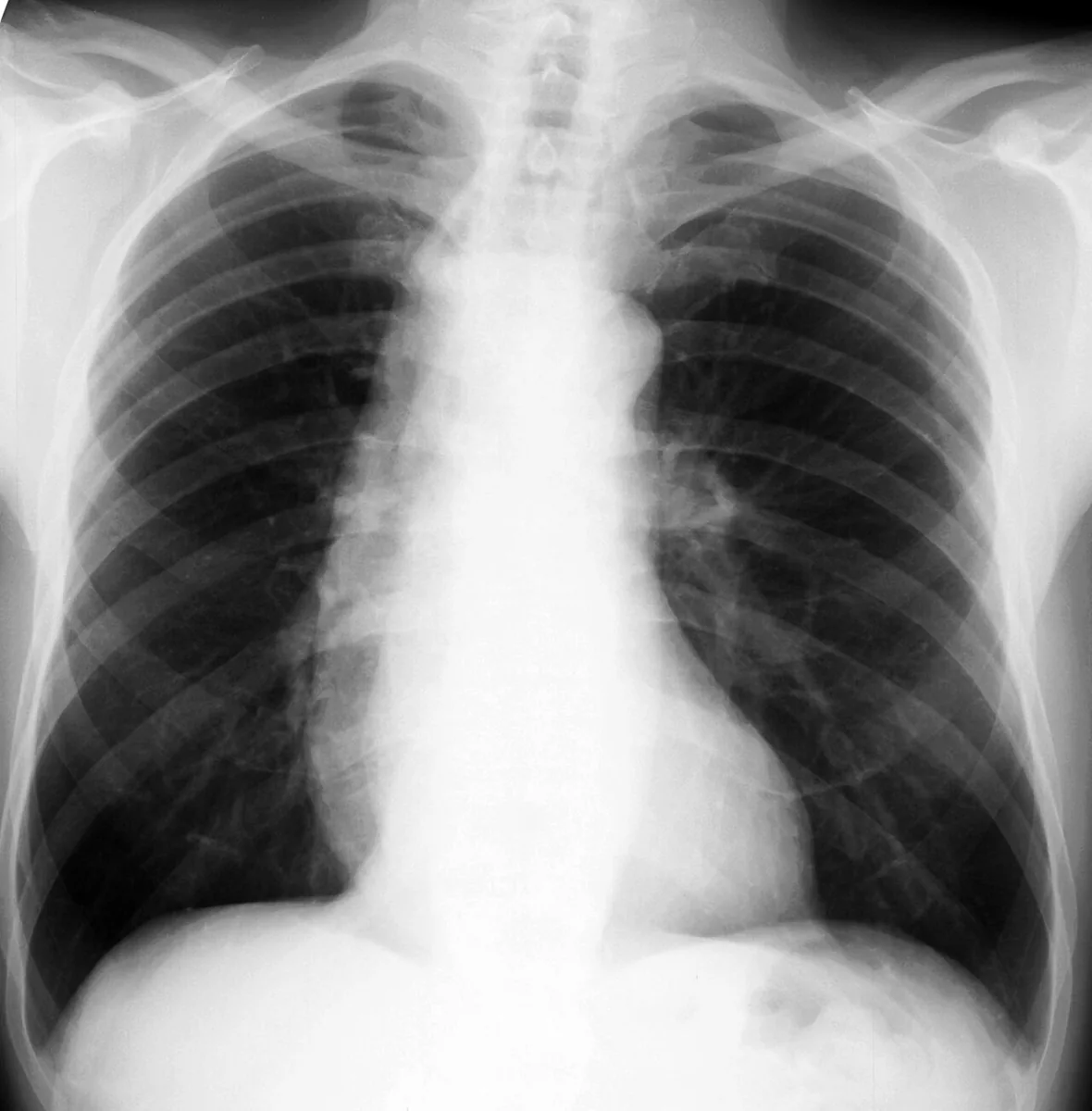 X-ray
X-ray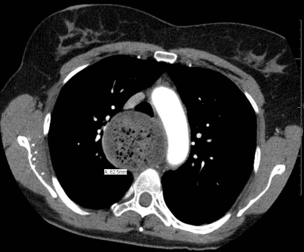 CT
CTDefinition
Achalasia is a neurogenic esophageal motility disorder with impaired peristalsis and absent relaxation of the lower esophageal sphincter on swallowing. The cardinal symptoms are slowly progressive dysphagia, usually for both liquids and solids, and regurgitation of undigested food.
Classification
High-resolution manometry distinguishes three subtypes:
- Type I (classic achalasia): swallowing produces no change in esophageal pressure.
- Type II: swallowing increases pressure throughout the esophagus.
- Type III (spastic achalasia): swallowing often produces lumen-obliterating contractions.
Esophagogastric junction outflow obstruction (raised median integrated relaxation pressure with evidence of peristalsis), which may be an early variant of achalasia, is to be distinguished.
Occurrence & epidemiology
Achalasia occurs at all ages but usually begins in adulthood; the mean age at diagnosis is over 50 years.
Aetiopathogenesis
- Loss of ganglion cells in the myenteric plexus of the esophagus with denervation of the esophageal muscle.
- Cause of denervation: unknown; viral and autoimmune mechanisms are suspected.
- Tumors can cause achalasia by direct obstruction or as a paraneoplastic process (pseudo-achalasia).
- Chagas disease: destroys autonomic ganglia and can thereby cause achalasia.
- Consequence: increased pressure in the lower esophageal sphincter causes obstruction with secondary dilatation of the esophagus; undigested food and liquid are often retained in the esophagus.
Clinical features
- Onset: insidious, progressing over months to years.
- Dysphagia for both solids and liquids as the main symptom.
- Heartburn in up to about 40 % of patients.
- Nocturnal regurgitation of undigested food with cough and aspiration; aspiration pneumonia or, chronically, bronchiectasis can result.
- Chest pain: less common, with swallowing or spontaneous.
- Loss of weight: usually mild to moderate. Marked loss of weight, particularly in older people with rapidly developing dysphagia, suggests pseudo-achalasia due to a tumor at the esophagogastric junction.
Diagnosis
- High-resolution manometry: the preferred test; incomplete relaxation of the lower esophageal sphincter with raised median integrated relaxation pressure and complete absence of peristalsis; classification into types I–III.
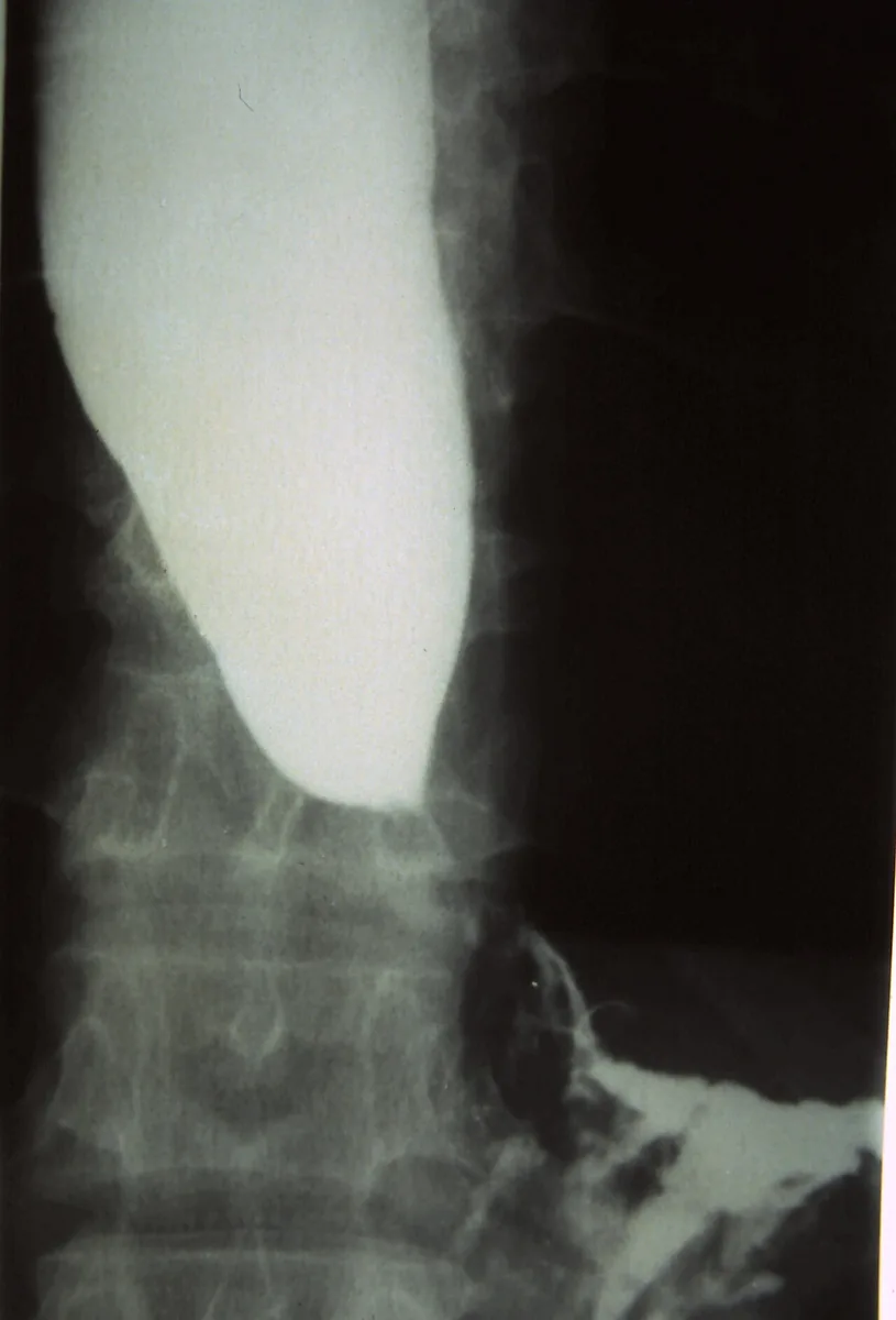
- Barium swallow: absent progressive peristalsis, an often greatly dilated esophagus with a beak-like narrowing at the lower sphincter ("bird's beak").
- Esophagoscopy: dilatation and chronic stasis changes of the mucosa without an obstructing lesion; the lower sphincter may be closed, and a typical "pop" is often felt as the endoscope enters the stomach.
- Impedance planimetry (FLIP): measures cross-sectional area and distensibility of the esophagogastric junction; helpful when manometry is equivocal despite high suspicion.
- If pseudo-achalasia is suspected: CT of the chest and abdomen or endoscopic ultrasound with biopsy.
Keep learning in the app
Further reading (open access)
Cross-references
More topics: Gastroenterology
Note: Learning content for medical education – not a treatment recommendation and no substitute for diagnosis or treatment decisions in individual cases. Treatment and management are deliberately not covered on this page.