Celiac disease
Board exam relevance: in 7 of 105 exam reports · rank 53- Synonyms
- coeliac disease, gluten intolerance, celiac sprue, gluten-sensitive enteropathy, non-tropical sprue
- Specialty
- Internal medicine · Gastroenterology
- Images
- Endoscopy 1 · Histology 2
- Last updated
- 10/2026 · Dr. Pascal Bafteh
Contents
Images (3)
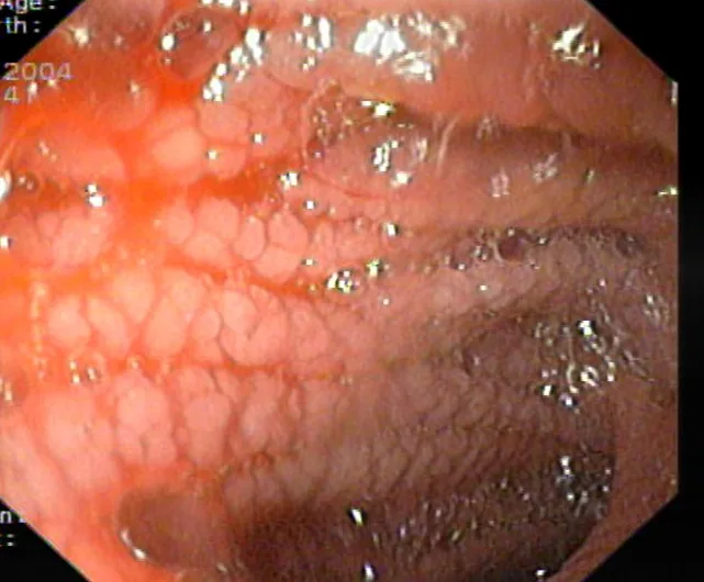 Endoscopy
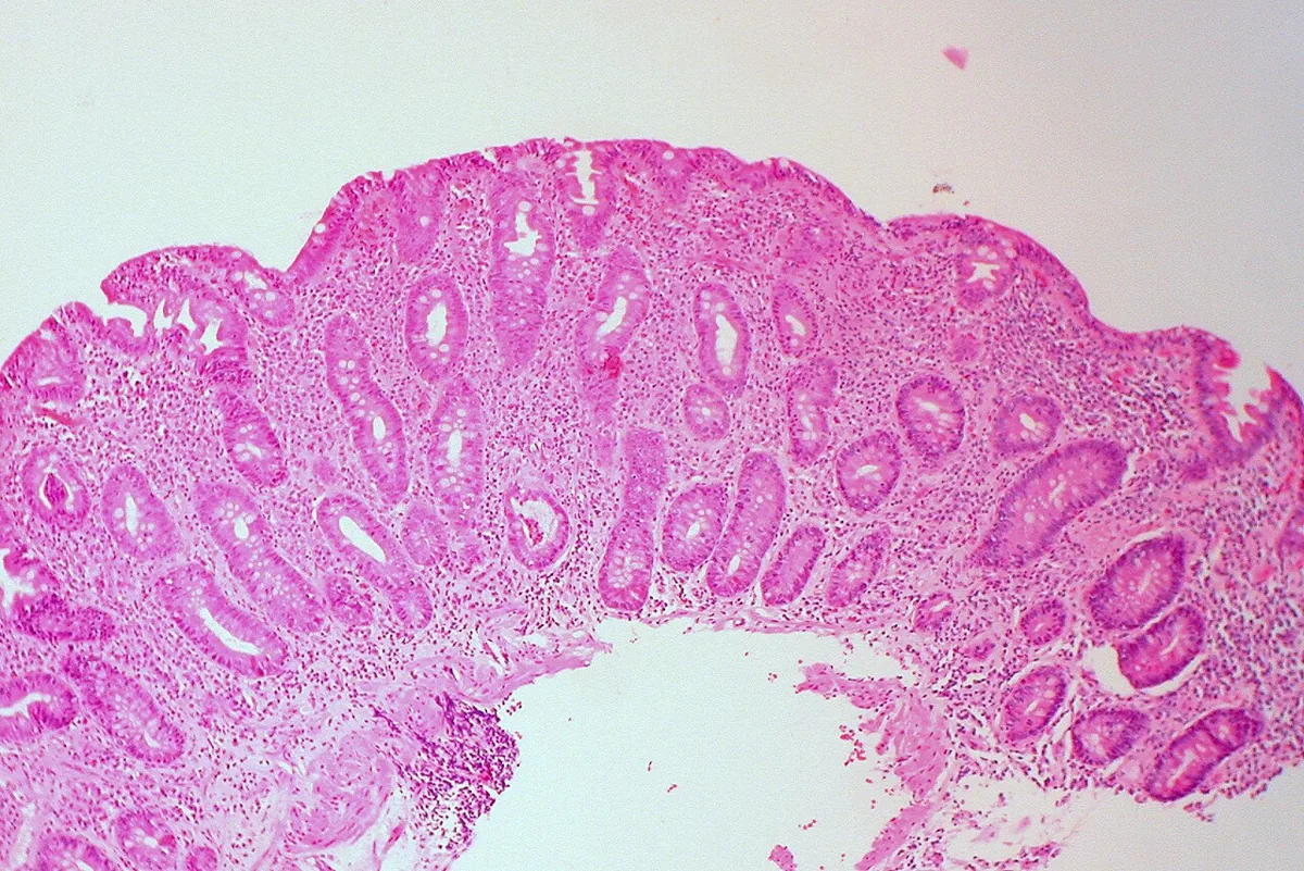 Histology
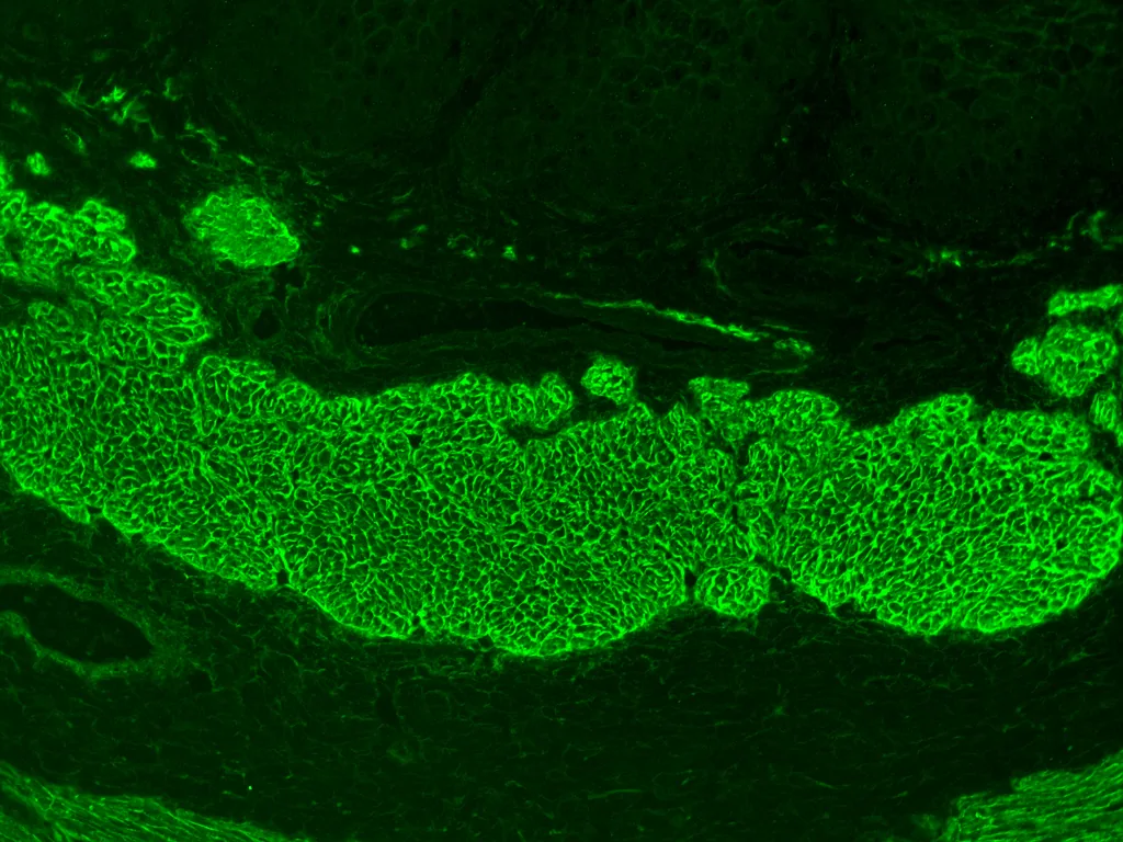 Histology
Definition
Celiac disease is an immunologically mediated disease in genetically susceptible people, caused by intolerance to gluten. It results in inflammation of the small-bowel mucosa with villous atrophy and consequently malabsorption.
Occurrence & epidemiology
Based on serologic screening of blood donors, celiac disease may affect up to 1.4% of the global population; the prevalence of biopsy-proven celiac disease is about half that and ranges from 0.4 to 0.8% depending on region. About 7.5% of first-degree relatives are affected, with a higher prevalence in females than males. Onset is generally in childhood but may occur later. People with lymphocytic colitis, Down syndrome, type 1 diabetes mellitus and Hashimoto thyroiditis are at increased risk.
Aetiopathogenesis
Celiac disease is a hereditary disorder caused by sensitivity to the gliadin fraction of gluten, a protein found in wheat; similar proteins are present in rye and barley. In genetically susceptible people, gluten-sensitive T cells are activated when gluten-derived peptide epitopes are presented. The resulting inflammatory response causes the characteristic villous atrophy in the small bowel. More than 95% of patients carry the HLA-DQ2 or HLA-DQ8 haplotype, although these haplotypes are not specific for celiac disease.
Clinical features
The clinical presentation varies so widely that no typical presentation exists; some patients are asymptomatic or have only signs of nutritional deficiency.
- Infants and young children (after introduction of cereals): failure to thrive, apathy, anorexia, pallor, generalized hypotonia, abdominal distention, muscle wasting; soft, bulky, clay-colored, foul-smelling stools.
- Older children: anemia or failure to grow normally.
- Adults: most commonly lassitude, weakness and anorexia; sometimes mild, intermittent diarrhea; steatorrhea of varying severity; weight loss, rarely to the point of being underweight.
- Signs of deficiency: anemia, glossitis, angular stomatitis, aphthous ulcers; consequences of vitamin D and calcium deficiency such as osteomalacia, osteopenia and osteoporosis.
- Fertility: reduced in both men and women, sometimes amenorrhea in women.
- Dermatitis herpetiformis: in about 17%; an intensely pruritic papulovesicular rash, symmetrically distributed over the extensor surfaces of the elbows and knees, the buttocks, shoulders and scalp.
Histology
Duodenal biopsies (including the second portion of the duodenum) show shortened or absent villi (villous atrophy), increased intraepithelial cells and crypt hyperplasia. These findings are characteristic but not specific.
Biopsy and Marsh classification
- Biopsies: at least 6 samples from different parts of the duodenum, 2 of them from the duodenal bulb; villous atrophy may be confined to the bulb.
- Modified Marsh-Oberhuber classification: type 0 normal (no more than 25 intraepithelial lymphocytes per 100 epithelial cells); type 1 more than 25 intraepithelial lymphocytes per 100 epithelial cells with normal crypts and villi; type 2 additionally crypt hyperplasia; types 3a, 3b and 3c additionally mild to moderate, subtotal or total villous atrophy.
Diagnosis
- Suspicion: clinically and with laboratory evidence of malabsorption; a family history is a valuable clue. Celiac disease is a consideration in iron deficiency without obvious gastrointestinal bleeding.
- Serology: tissue transglutaminase IgA (tTG-IgA) and endomysial IgA antibodies (EMA), with sensitivity and specificity > 90%. If either test is positive, a small-bowel biopsy follows; if both are negative, celiac disease is extremely unlikely.
- Prerequisites: all serologic tests are performed while gluten is being consumed. Serum IgA is measured at the same time, because IgA deficiency can cause false-negative results; with IgA deficiency, IgG antibodies against tTG and deamidated gliadin peptide (DGP) are measured.
- Small-bowel biopsy: for confirmation, from the second portion of the duodenum.
- HLA typing: absence of HLA-DQ2 and -DQ8 effectively rules out celiac disease; helpful when biopsy and serology are discordant.
- Other laboratory findings: anemia (iron-deficiency anemia in children, folate-deficiency anemia in adults), hypoalbuminemia, hypocalcemia, hypokalemia, hyponatremia, elevated alkaline phosphatase, prolonged prothrombin time.
Children and adolescents
- Diagnosis without biopsy: possible in children and adolescents under 18 years if tTG-IgA reaches at least 10 times the cut-off (quality-assured test) and EMA is positive in a second, independent blood sample.
- Adults: confirmation usually by duodenal biopsies; omitting them is considered only in exceptional cases, e.g. with tTG-IgA above 10 times the cut-off when upper endoscopy is not possible.
Keep learning in the app
Further reading (open access)
Cross-references
More topics: Gastroenterology
Note: Learning content for medical education – not a treatment recommendation and no substitute for diagnosis or treatment decisions in individual cases. Treatment and management are deliberately not covered on this page.