Upper gastrointestinal bleeding
Board exam relevance: in 5 of 105 exam reports · rank 69- Synonyms
- UGIB, upper GI bleed, vomiting blood, haematemesis, melaena, black tarry stool, bleeding ulcer
- Specialty
- Internal medicine · Gastroenterology
- Images
- Endoscopy 1 · Clinical 1
- Last updated
- 10/2026 · Dr. Pascal Bafteh
Contents
Images (2)
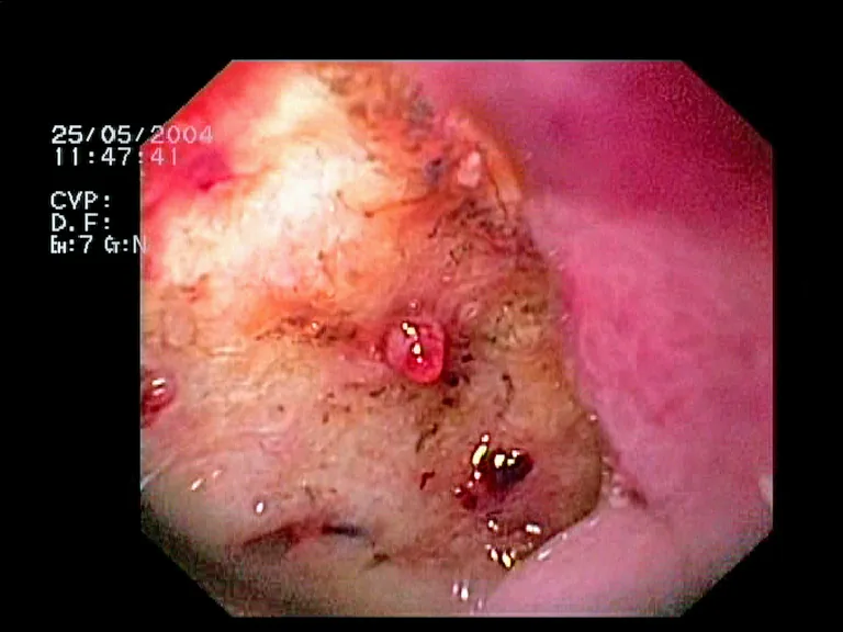 Endoscopy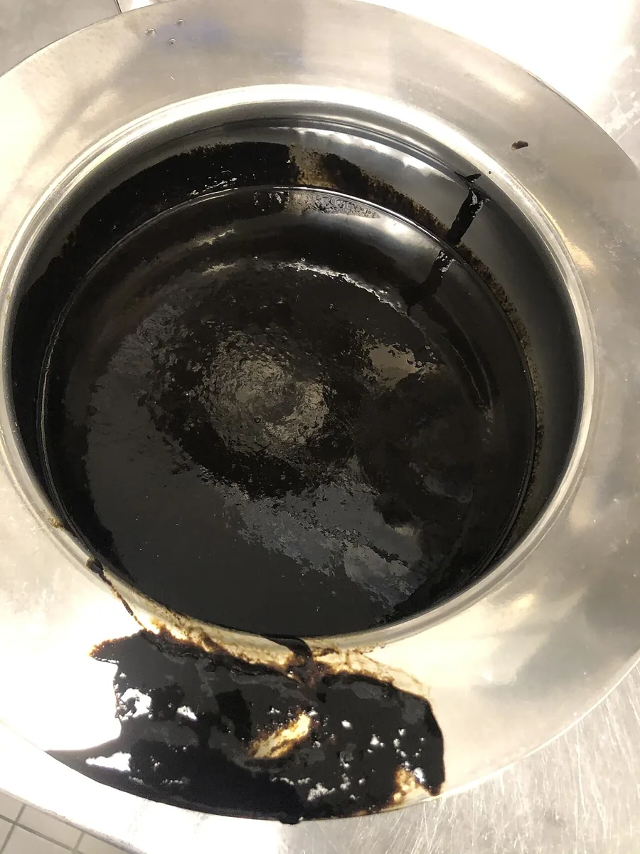
Definition
Upper gastrointestinal bleeding is bleeding above the ligament of Treitz, i.e. from the esophagus, stomach or duodenum. Gastrointestinal bleeding can be overt or occult; its manifestations depend on the location and rate of bleeding. Variceal and non-variceal bleeding are distinguished.
Classification
Forrest classification (endoscopic appearance of bleeding ulcers, introduced in 1974):
- Ia: spurting bleeding
- Ib: oozing bleeding
- IIa: non-bleeding visible vessel
- IIb: adherent clot
- IIc: pigmented base
- III: clean base
Forrest Ia and Ib are independent risk factors for persistent or recurrent bleeding.
Risk scores:
- Glasgow–Blatchford score (before endoscopy, 0–23 points): hemoglobin, urea, heart rate, systolic blood pressure, comorbidities (liver disease, heart failure) and presenting features (syncope, melaena).
- Rockall score (0–11 points): age, hemodynamic status, comorbidities, endoscopic diagnosis and stigmata of recent hemorrhage; a version without endoscopic items can be used before endoscopy.
- ABC score: age, urea, albumin, creatinine, altered mental status, cirrhosis, disseminated cancer and American Society of Anesthesiologists classification; for upper and lower bleeding.
Aetiopathogenesis
Common causes (share of upper gastrointestinal bleeds):
- Duodenal ulcer (15–29 %) and gastric ulcer (14–16 %); gastric and duodenal ulcers together account for up to 50 %.
- Varices (5–33 %).
- Mallory–Weiss tear (6–15 %).
- Gastric or duodenal erosions (3–15 %) and erosive esophagitis (2–15 %).
- Angiomas (1–5 %), gastrointestinal stromal tumors (1–5 %), arteriovenous malformations (< 5 %), hemobilia.
- Other vascular lesions: Dieulafoy lesion (an abnormally large artery, mainly in the proximal stomach, with possible massive bleeding), gastric antral vascular ectasia ("watermelon stomach"), hereditary hemorrhagic telangiectasia.
Predisposing factors: chronic liver disease, hereditary coagulation disorders and drugs that affect coagulation or platelet function (e.g. NSAIDs, certain SSRI-type antidepressants) or weaken mucosal defences.
Clinical features
- Hematemesis: vomiting of red blood; indicates upper bleeding, usually from an ulcer, vascular lesion or varix.
- Coffee-ground vomitus: dark brown, granular vomitus; bleeding has slowed or stopped, and hemoglobin has been oxidised to brown hematin by gastric acid.
- Melaena: black, tarry stool; usually upper bleeding, but also possible from the small bowel or right colon. About 100–200 ml of blood in the upper gastrointestinal tract is required; melaena may persist for several days after bleeding has stopped.
- Hematochezia: passage of gross blood per rectum; usually lower bleeding, but possible with vigorous upper bleeding and rapid transit.
- Hypovolaemia and shock: syncope, hypotension, pallor, sweating, tachycardia, oliguria; orthostatic hypotension (a fall of > 20 mmHg systolic or 10 mmHg diastolic with a rise in heart rate) often develops after acute loss of more than 2 units of blood.
- Consequences in pre-existing disease: angina or myocardial infarction in ischemic heart disease; hepatic encephalopathy or hepatorenal syndrome in liver disease.
- Clues to the cause: epigastric pain relieved by food (ulcer, although many bleeding ulcers are painless); cirrhosis or stigmata of liver disease (varices); vomiting and retching before bleeding (Mallory–Weiss); dysphagia (esophageal cancer, stricture); loss of weight (tumor).
Diagnosis
- History: quantity and frequency of blood loss, blood with the first vomit or only after non-bloody vomiting, distinction from hemoptysis; previous bleeding, liver disease, bleeding disorders, drugs and alcohol.
- Physical findings: vital signs and signs of shock; signs of bleeding disorders (petechiae, ecchymoses), chronic liver disease and portal hypertension; digital rectal examination (stool color, masses, fissures).
- Laboratory tests: full blood count; with more significant bleeding, coagulation (platelets, prothrombin time/INR, PTT) and liver function tests; blood typing and cross-match if bleeding continues. A blood urea nitrogen to creatinine ratio > 30 suggests an upper source.
- Nasogastric tube: bloody aspirate indicates active upper bleeding; however, up to 15 % of upper bleeds show no blood in the aspirate.
- Upper endoscopy: the central investigation for locating and characterising the bleeding source.
- Barium studies: no role in acute bleeding.
- Angiography: in selected cases of upper and lower bleeding.
Keep learning in the app
Further reading (open access)
Cross-references
More topics: Gastroenterology
Note: Learning content for medical education – not a treatment recommendation and no substitute for diagnosis or treatment decisions in individual cases. Treatment and management are deliberately not covered on this page.