Lower gastrointestinal bleeding
Board exam relevance: in 4 of 105 exam reports · rank 90- Synonyms
- LGIB, lower GI bleed, rectal bleeding, blood in the stool, haematochezia, diverticular bleeding, bowel bleeding
- Specialty
- Internal medicine · Gastroenterology
- Images
- CT 2
- Last updated
- 10/2026 · Dr. Pascal Bafteh
Contents
Images (2)
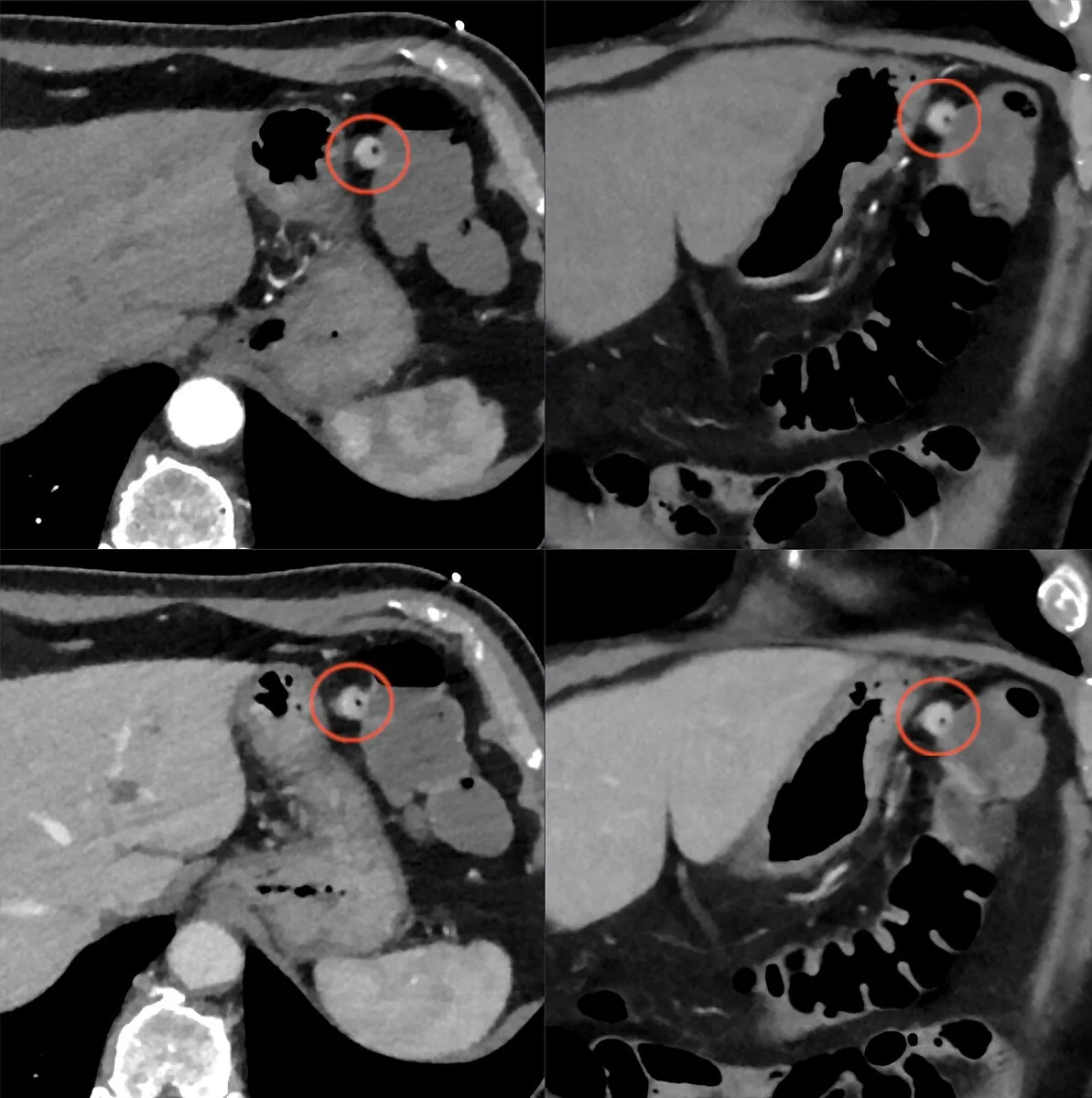 CT
CT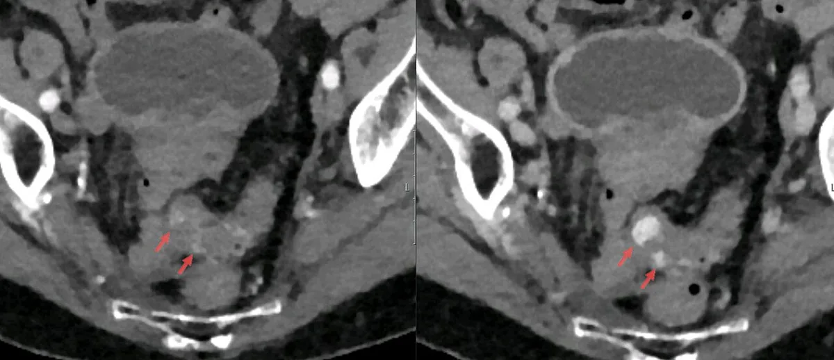 CT
CTDefinition
Lower gastrointestinal bleeding was traditionally defined as bleeding from a source distal to the ligament of Treitz, including the small and large bowel. With the spread of capsule endoscopy and balloon-assisted enteroscopy, three categories are increasingly distinguished: upper, mid (small bowel) and lower gastrointestinal bleeding. The cardinal symptom is hematochezia, the passage of gross blood per rectum.
Occurrence & epidemiology
Acute lower gastrointestinal bleeding has increased with the ageing population and the growing use of antithrombotic drugs; the rate of bleeds requiring hospital care is estimated at 33–87 per 100,000 population. Most bleeds stop spontaneously. Diverticular bleeding and angiodysplasia, however, tend to cause continuous or recurrent bleeding.
Aetiopathogenesis
Causes of acute lower bleeding in Western countries (proportions):
- Diverticular bleeding (30–65 %): the most common cause in Western countries; occurs in 10–15 % of people with diverticulosis. Diverticula form at weak points of the muscular layer next to intramural blood vessels; the source of bleeding is an arteriole. The right colon is the source in more than 50 %; NSAIDs increase the risk.
- Ischemic colitis (5–20 %) and hemorrhoids (5–20 %).
- Colorectal polyps and neoplasms (2–15 %).
- Angiodysplasia (5–10 %): dilated, tortuous vessels, typically in the caecum and ascending colon; mainly in people over 60 and an important cause of lower bleeding in this age group; associated with renal failure, aortic stenosis, cirrhosis and CREST syndrome.
- Other causes: post-polypectomy bleeding (2–7 %), inflammatory bowel disease (3–5 %), infectious colitis (2–5 %), rectal ulcer, colorectal varices, proctitis after exposure to ionising rays, drug-induced colitis, Dieulafoy lesion, anal fissures, Meckel's diverticulum.
- Upper source: brisk upper bleeding can also cause hematochezia.
Clinical features
- Diverticular bleeding: painless hematochezia with fresh blood or maroon stool; blood loss is usually moderate to severe, and right-sided bleeding rarely presents as melaena.
- Angiodysplasia: painless, often intermittent bleeding with occult blood in the stool or modest amounts of bright red blood; major bleeding is unusual.
- Colitis (ischemic, infectious, inflammatory): often diarrhea, abdominal pain and tenderness.
- Hemorrhoids and fissures: fresh blood only on the toilet paper or the surface of formed stool; blood mixed with stool indicates a more proximal source.
- Tumors: loss of weight and altered bowel habit; occult blood may be the first sign of colon cancer or a polyp.
- Circulation: syncope, hypotension, tachycardia; hematochezia with hemodynamic instability may also indicate brisk upper bleeding.
Diagnosis
- History and physical findings: comorbidities, drugs, symptoms, vital signs; digital rectal examination and anoscopy (hemorrhoids).
- Excluding an upper source: a blood urea nitrogen to creatinine ratio > 30 and a bloody or coffee-ground nasogastric aspirate point to upper bleeding; upper endoscopy is then performed.
- Laboratory tests: full blood count, coagulation, liver function tests; blood typing and cross-match if bleeding continues.
- Colonoscopy: in hematochezia (unless clearly hemorrhoidal) after bowel preparation; with suspected hemorrhoids, sigmoidoscopy and anoscopy are often sufficient.
- CT angiography: the first investigation in ongoing, hemodynamically significant hematochezia; more sensitive than catheter angiography and detects bleeding from about 0.3 ml/min.
- Catheter angiography: requires bleeding rates above 0.5 ml/min; labelled red blood cell scintigraphy when CT angiography is not possible.
- Occult bleeding with negative upper endoscopy and colonoscopy: small bowel work-up with CT enterography, enteroscopy, capsule endoscopy or scintigraphy.
Keep learning in the app
Further reading (open access)
Cross-references
More topics: Gastroenterology
Note: Learning content for medical education – not a treatment recommendation and no substitute for diagnosis or treatment decisions in individual cases. Treatment and management are deliberately not covered on this page.