Influenza (flu)
Board exam relevance: in 4 of 105 exam reports · rank 90- Synonyms
- flu, seasonal influenza, influenza A, influenza B
- Specialty
- Internal medicine · Infectious diseases
- Images
- X-ray 1 · Gross specimen 1
- Last updated
- 10/2026 · Dr. Pascal Bafteh
Contents
Images (2)
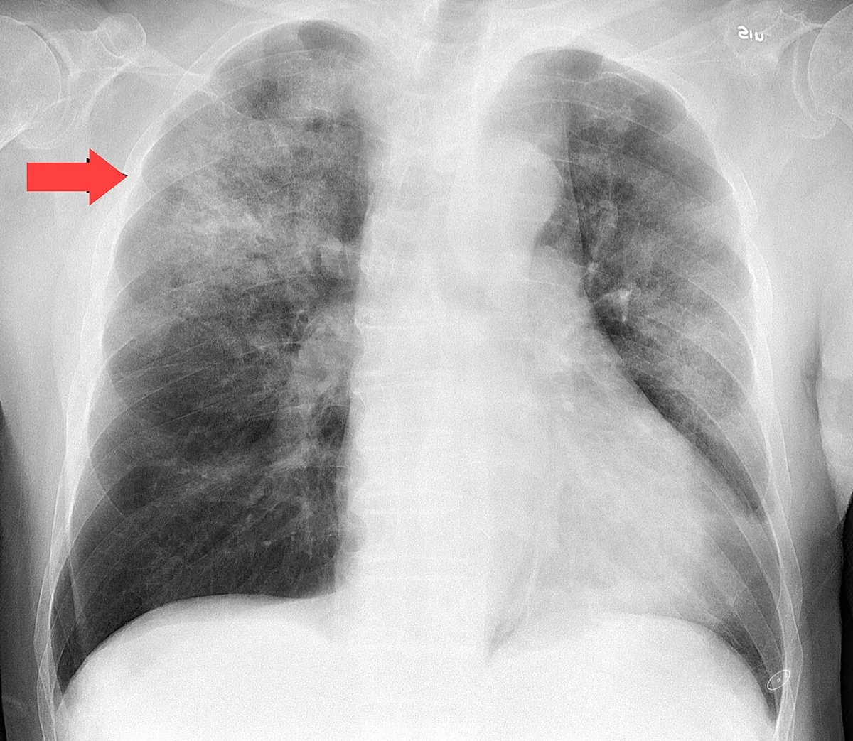 X-ray
X-ray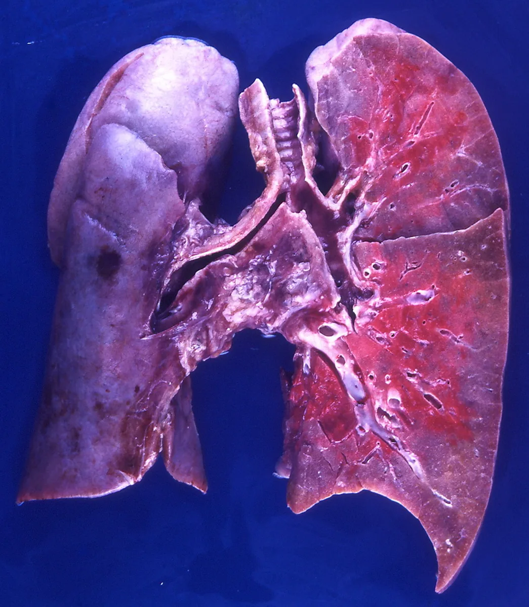 Gross specimen
Definition
Influenza ("the flu") is an acute respiratory infection caused by influenza viruses that occurs in annual epidemics. It is characterized by sudden onset with fever, cough or sore throat, and muscle aches and headache. It is distinct from the common cold caused by other respiratory viruses.
Classification
- Influenza A: subtypes according to the surface proteins hemagglutinin (H) and neuraminidase (N), e.g. A(H3N2); since 2009, A(H1N1)pdm09 and A(H3N2) have circulated in humans.
- Influenza B: no subtypes but lineages; since 2020 only the Victoria lineage has been detected.
- Influenza C: of little relevance for humans.
- Seasonal, zoonotic and pandemic influenza: novel viruses resulting from reassortment of gene segments (antigenic shift) can cause pandemics.
Occurrence & epidemiology
- Worldwide distribution; in temperate zones epidemics occur in the winter months, in the tropics year-round.
- In Germany, activity usually rises markedly in January or February; an epidemic lasts 3–4 months on average. An estimated 5–20 % of the population is infected per season; the RKI estimates 1–7 million influenza-related physician visits per year.
- Physician visits involve all age groups; hospital stays are most frequent in young children and older adults.
Aetiopathogenesis
- Pathogen: orthomyxoviruses with an RNA genome divided into eight segments and the surface proteins hemagglutinin and neuraminidase.
- Antigenic drift: ongoing point mutations that allow the virus to evade strain-specific immunity; the basis of annual epidemics.
- Antigenic shift: reassortment of gene segments, e.g. between human and avian or swine viruses; wild waterfowl are the reservoir of nonhuman influenza A viruses.
- Reservoir of seasonal influenza: humans only.
- Transmission: mainly via exhaled virus-containing droplets and small particles at close range, especially when coughing and sneezing; also via hands and contaminated surfaces.
- Incubation period: short, about 1–3 days (range up to 4 days, mean 48 hours).
- Infectiousness: viral shedding on average about 4–5 days from symptom onset, longer in children and the severely ill; possible even before symptoms begin.
Clinical features
Symptoms
- Typical course: sudden onset with fever, chills, marked prostration, headache (often with photophobia and retrobulbar pain), aching limbs and back pain, dry cough, sore throat and substernal burning; sometimes rhinorrhea, sweating, rarely nausea, vomiting and diarrhea.
- Spectrum: about one third of infections are febrile, one third milder and one third asymptomatic.
- Duration: usually 5–7 days; cough and malaise may persist for more than 2 weeks.
- Risk groups for severe disease: older adults, chronic heart and lung disease, metabolic disorders such as diabetes mellitus, immunodeficiency, neurologic and neuromuscular diseases, severe obesity and pregnancy (especially in advanced stages). Deterioration often occurs 3–10 days after symptom onset.
Complications
- Primary influenza pneumonia caused by the virus itself, up to acute respiratory distress syndrome (ARDS).
- Secondary bacterial infection (including pneumococci, staphylococci, Hemophilus influenzae), suspected with recurrent fever and cough after apparent improvement.
- Exacerbation of chronic lung diseases such as asthma and COPD; myocardial infarction and heart failure in pre-existing heart disease.
- Other organs: myositis and rhabdomyolysis, myocarditis, pericarditis, encephalitis.
- Children: otitis media, febrile seizures, benign acute myositis (calf pain); Reye syndrome with encephalopathy and fatty liver, especially after ingestion of salicylates.
Diagnosis
- Clinical diagnosis: during an influenza epidemic, the typical symptoms have good predictive value. Other respiratory pathogens (e.g. rhinoviruses, RSV, metapneumoviruses, mycoplasmas) can cause similar pictures; reliable distinction is possible only by laboratory testing. In older or immunocompromised people, symptoms may be nonspecific and without fever.
- PCR (RT-PCR) of nasal or throat swab: gold standard for sensitivity and specificity; distinguishes types and subtypes. Multiplex assays detect SARS-CoV-2 and RSV at the same time.
- Rapid antigen tests: result in 10–15 minutes, high specificity but only low to moderate sensitivity; a negative result does not exclude influenza. Rapid molecular tests are considerably more sensitive.
- Other methods: immunofluorescence, viral culture and serology (rise in titer between acute and convalescent serum, mainly for epidemiologic purposes).
- With lower respiratory tract involvement: pulse oximetry and chest X-ray. Primary influenza pneumonia shows focal or diffuse interstitial infiltrates up to an ARDS pattern; secondary bacterial pneumonia more often lobar or segmental opacities.
Keep learning in the app
Further reading (open access)
Cross-references
More topics: Infectious diseases
Note: Learning content for medical education – not a treatment recommendation and no substitute for diagnosis or treatment decisions in individual cases. Treatment and management are deliberately not covered on this page.