Sepsis and septic shock
Board exam relevance: in 10 of 105 exam reports · rank 27- Synonyms
- blood poisoning, septicemia, Sepsis-3
- Specialty
- Internal medicine · Infectious diseases
- Images
- Clinical 1 · Blood smear & cytology 1 · Gross specimen 1
- Last updated
- 10/2026 · Dr. Pascal Bafteh
Contents
Images (3)
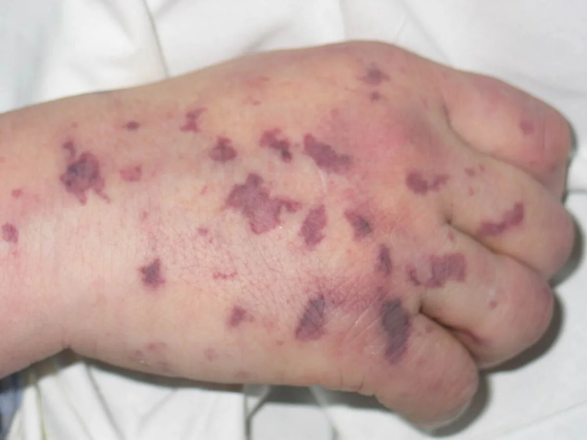
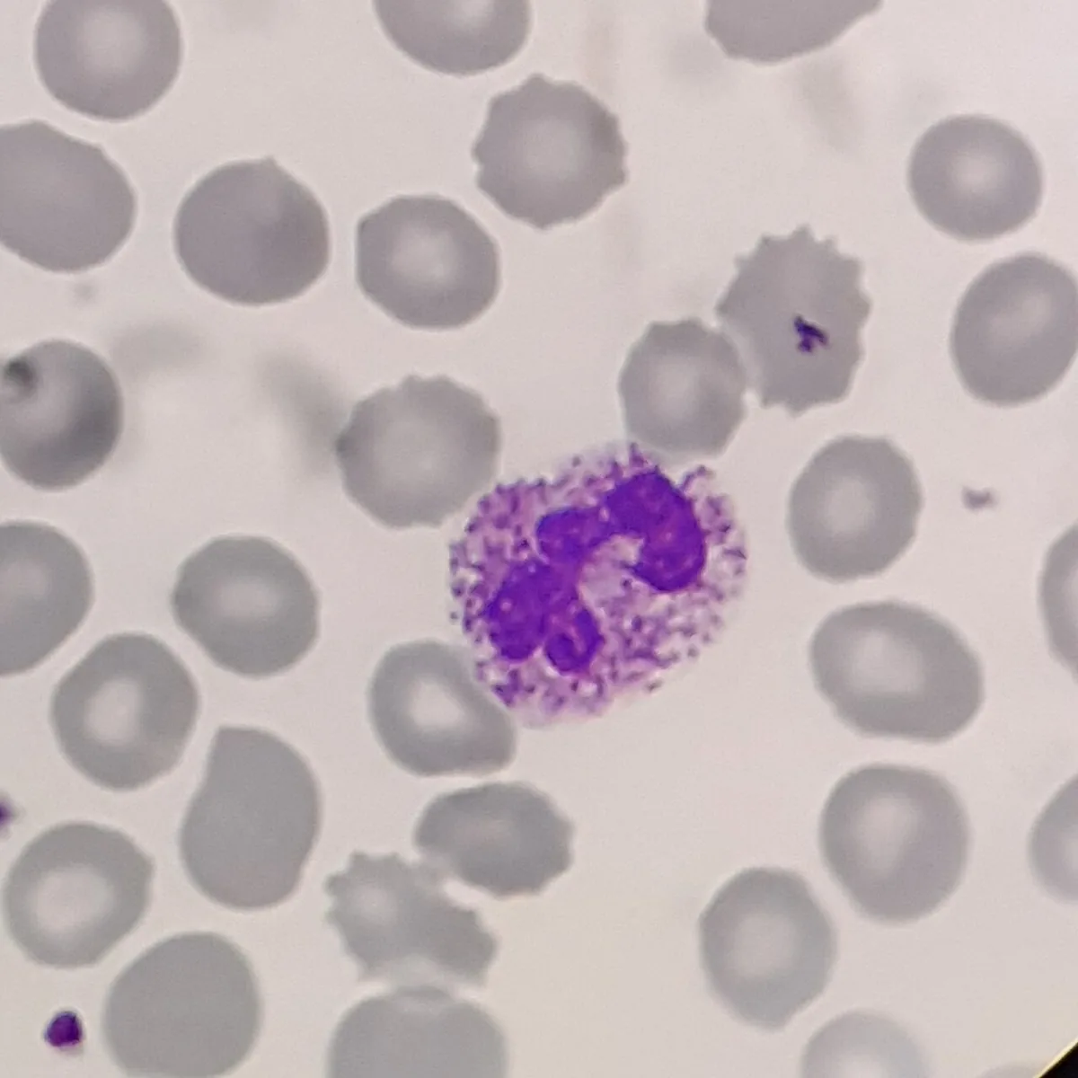 Blood smear & cytology
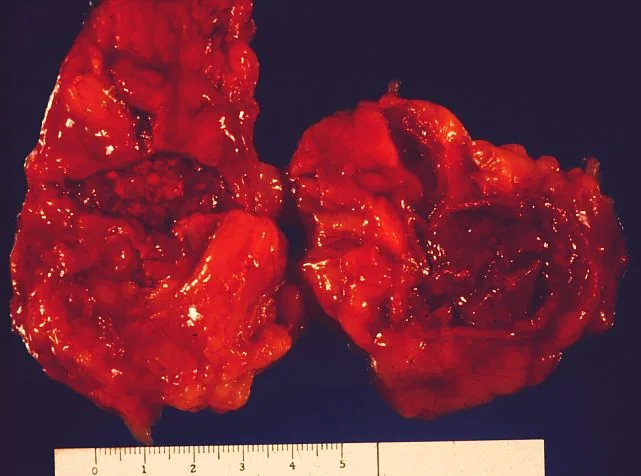 Gross specimen
Definition
Sepsis is defined by the current consensus definition (Sepsis-3, 2016) as life-threatening organ dysfunction caused by a dysregulated host response to infection. Clinically, organ dysfunction is identified as an acute increase in the total SOFA score of at least 2 points consequent to the infection.
Septic shock is a subset of sepsis with profound circulatory and cellular/metabolic abnormalities. It is present when, despite adequate intravascular volume, persistent hypotension requires vasoactive agents to maintain a mean arterial pressure (MAP) of at least 65 mm Hg and serum lactate is above 2 mmol/L.
Classification
- By place of acquisition: community-acquired sepsis and sepsis acquired in the course of health care (nosocomial).
- By source: pulmonary, intra-abdominal, genitourinary, postoperative, arising from indwelling foreign material (e.g. vascular or urinary catheters); occasionally the source remains occult.
Occurrence & epidemiology
- According to World Health Organization data (published 2020), about 48.9 million sepsis cases occurred worldwide per year; almost half (around 20 million) affected children under 5 years of age.
- Of every 1,000 patients in hospital, an estimated 15 develop sepsis as a complication of health care.
Aetiopathogenesis
- Pathogens: mostly gram-negative bacilli or gram-positive cocci; less often Candida and other fungi or viruses. In immunocompromised patients, uncommon bacteria or fungi occur.
- Most common sources of infection: lungs, abdomen and genitourinary tract; after recent surgery also a postoperative infection.
- Risk factors: diabetes mellitus; liver disease including cirrhosis and MASLD; leukopenia and neutropenia (especially with cancer or cytotoxic medicines); indwelling foreign material such as endotracheal tubes, vascular and urinary catheters; use of immunosuppressive drugs; recent hospital stay; HIV infection and other forms of immunodeficiency; chronic kidney disease; pregnancy.
Pathophysiology
An inflammatory stimulus, such as a bacterial toxin, triggers the production of cytokines such as TNF and interleukin-1; they promote adhesion of neutrophils to the endothelium, activate coagulation and generate microthrombi.
Initially, arteries and arterioles dilate, peripheral resistance falls and cardiac output usually rises (warm phase). Later, cardiac output falls and signs of hypoperfusion appear (cold phase).
Shunting past the exchange capillaries (distributive defect) and microthrombi reduce oxygen supply to the tissues, leading to organ dysfunction. Consumption of clotting factors and excessive fibrinolysis can cause coagulopathy.
Clinical features
Symptoms
- General signs: fever (or hypothermia), chills, tachycardia, tachypnea, sweating and marked malaise; plus symptoms of the underlying infection, e.g. cough in pneumonia.
- Nonspecific presentation: signs may be subtle and resemble other disorders. Older adults often mount only a weak inflammatory response.
- Early warning sign: confusion or decreased alertness, especially in the very young and the very old.
Septic shock
- Warm phase: falling blood pressure with paradoxically warm skin.
- Cold phase: cool, pale extremities, peripheral cyanosis and mottled skin; prolonged capillary refill time.
Organ dysfunction
- Brain: confusion and decreased alertness.
- Lungs: hypoxemic respiratory failure up to acute respiratory distress syndrome (ARDS) with diffuse infiltrates.
- Kidneys: oliguria and rising urea and creatinine as signs of renal dysfunction.
- Liver: rising bilirubin and transaminases.
- Coagulation: thrombocytopenia, disseminated intravascular coagulation.
- Heart and metabolism: arrhythmias, lactic acidosis.
Diagnosis
Clinical scores
- qSOFA (quick SOFA): 1 point each for respiratory rate of 22/min or higher, altered mentation and systolic blood pressure of 100 mm Hg or lower. A score of 2 or more indicates increased risk in suspected infection; the score requires no laboratory values.
- SOFA score: six organ systems scored 0 to 4 points each: respiration (PaO2/FiO2 ratio), coagulation (platelets, 1 point below 150 × 10⁹/L), liver (bilirubin), cardiovascular (MAP below 70 mm Hg or need for vasoactive agents), CNS (Glasgow Coma Scale) and kidney (creatinine or urine output).
- SIRS criteria (historical, not part of the definition): at least 2 of 4: temperature above 38 °C or below 36 °C; respiratory rate above 20/min or PaCO2 below 32 mm Hg; heart rate above 90/min; white blood cell count above 12,000/µL, below 4,000/µL or more than 10 % immature (band) forms.
Laboratory tests and microbiology
- Complete blood count with differential: white blood cells decreased (below 4,000/µL) or increased (above 15,000/µL). Thrombocytopenia.
- Lactate: elevated as a sign of tissue hypoxia; part of the definition of shock.
- Arterial blood gas analysis: early respiratory alkalosis from hyperventilation, later metabolic (lactic) acidosis; reduced PaO2/FiO2 ratio.
- Inflammatory markers: C-reactive protein and procalcitonin are often elevated but are not specific.
- Pathogen detection: blood cultures, urinalysis and urine culture, and cultures of further specimens depending on the suspected source (e.g. sputum, wound swab, cerebrospinal fluid, aspirates).
Imaging, ECG and echocardiography
- Imaging to locate the source: ultrasonography, chest X-ray, CT or MRI depending on the suspected source of infection; chest X-ray may show diffuse infiltrates in ARDS.
- ECG: hypoperfusion may cause nonspecific ST-T changes and arrhythmias even without infarction.
- Echocardiography: in septic shock, cardiac output is typically increased and peripheral vascular resistance decreased, whereas in most other forms of shock the reverse applies.
Keep learning in the app
Further reading (open access)
Cross-references
Note: Learning content for medical education – not a treatment recommendation and no substitute for diagnosis or treatment decisions in individual cases. Treatment and management are deliberately not covered on this page.