Bacterial meningitis
Board exam relevance: in 2 of 105 exam reports · rank 142- Synonyms
- brain fever, purulent meningitis, meningococcal meningitis, pneumococcal meningitis
- Specialty
- Internal medicine · Infectious diseases
- Images
- Gross specimen 1 · Clinical 1 · Blood smear & cytology 2
- Last updated
- 10/2026 · Dr. Pascal Bafteh
Contents
Images (4)
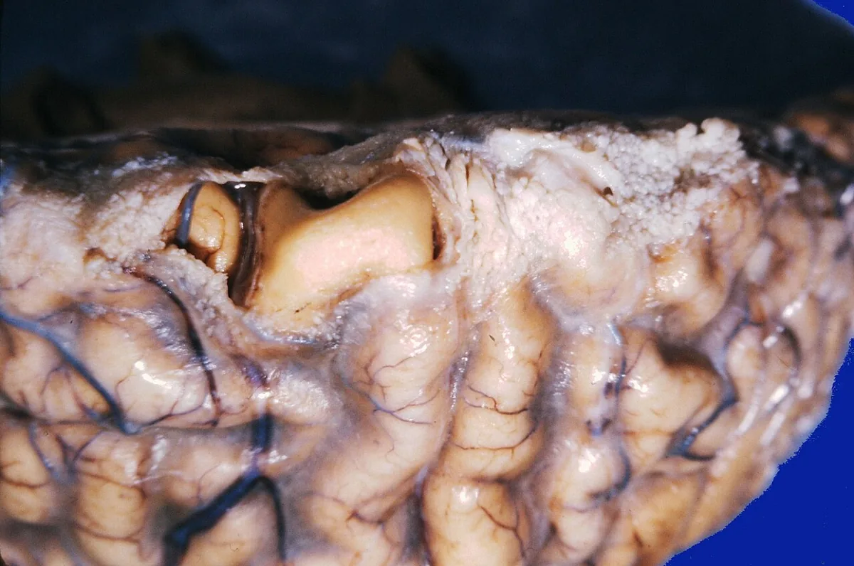 Gross specimen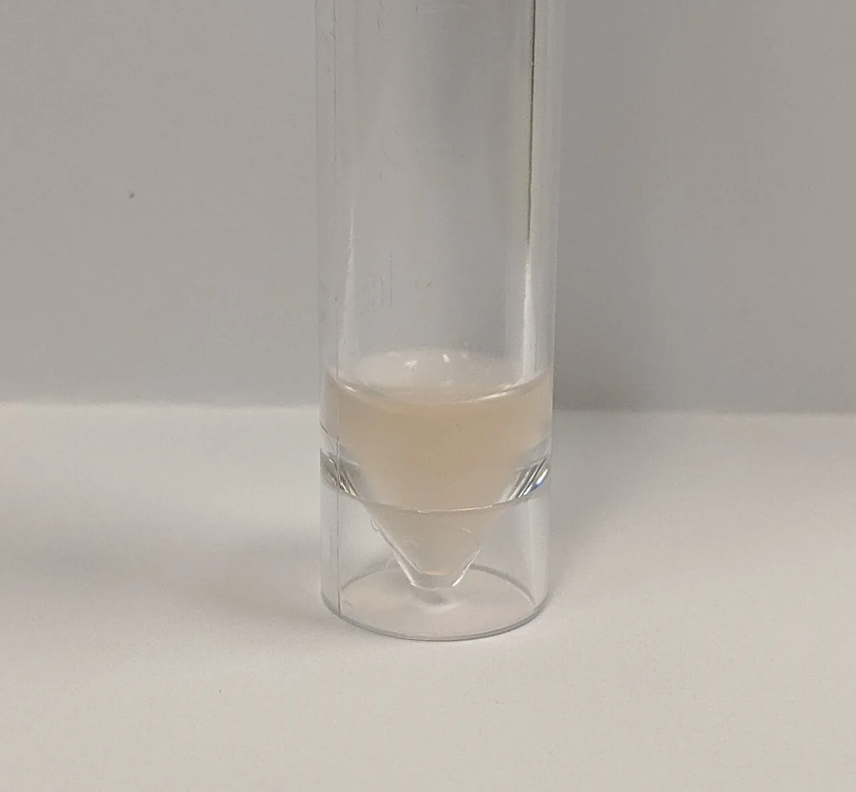
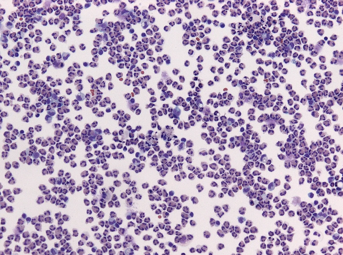 Blood smear & cytology
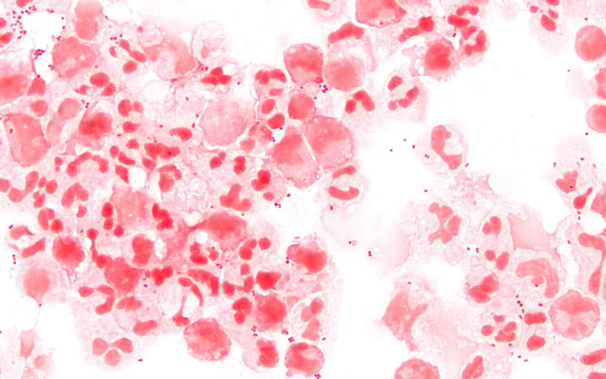 Blood smear & cytology
Definition
Acute bacterial meningitis is a rapidly progressive bacterial infection of the meninges and subarachnoid space. Leading symptoms are headache, fever and nuchal rigidity. Diagnosis is based on analysis of the cerebrospinal fluid. The disease is a medical emergency.
Classification
- Community-acquired meningitis: usually hematogenous, pathogens depending on age and immune status.
- Meningitis by contiguous spread: from the paranasal sinuses, middle ear or mastoid, sometimes with a CSF leak.
- Post-traumatic and postoperative meningitis: after penetrating head injury or after surgery on the brain or spine (e.g. infected ventricular shunt).
- Meningitis in immunodeficiency with a different pathogen spectrum.
Occurrence & epidemiology
- Hemophilus influenzae type b, formerly the most common cause of meningitis, is now rare in Western Europe.
- Invasive meningococcal disease in Germany: currently below 0.4 cases per 100,000 population per year, mostly serogroups B and Y; peak incidence in the 1st and 2nd year of life and a smaller peak at 15–19 years. About 10 % of the population carry meningococci asymptomatically in the nasopharynx.
Aetiopathogenesis
Pathogens
- Neonates and young infants: group B streptococci (S. agalactiae), E. coli and other gram-negative bacteria, Listeria monocytogenes.
- Older infants, children and young adults: Neisseria meningitidis and Streptococcus pneumoniae; Hemophilus influenzae type b mainly in regions where it is still common.
- Middle-aged and older adults: most commonly S. pneumoniae, less often N. meningitidis; with increasing age also Listeria and gram-negative bacteria.
- Staphylococcus aureus: possible at any age; the most common pathogen after penetrating head injury.
- After surgery on the brain or spine: including gram-negative bacteria (Klebsiella, Acinetobacter, E. coli, Pseudomonas) and staphylococci.
- Immunodeficiency: impaired cell-mediated immunity (advanced HIV infection, Hodgkin lymphoma, immunosuppressive drugs) favors Listeria and mycobacteria; impaired humoral immunity or splenectomy S. pneumoniae and N. meningitidis; neutropenia Pseudomonas aeruginosa and gram-negative enteric bacteria.
- Meningococci: gram-negative diplococci, transmitted by close contact with oropharyngeal secretions; incubation period usually 3–4 days (2–10 days).
Pathogenesis
Most pathogens colonize the nose and throat and are spread by droplets or throat secretions. They usually reach the meninges hematogenously, less often by contiguous spread from adjacent structures or through congenital or acquired defects of the skull and spine. Because cerebrospinal fluid normally contains few white blood cells, immunoglobulins and complement, bacteria initially multiply unhindered. Bacterial components then trigger an inflammatory response; CSF protein rises and CSF glucose falls owing to consumption and reduced transport. The brain parenchyma is often involved (cortical encephalitis, ventriculitis).
Clinical features
Symptoms
- Onset: usually 3–5 days of progressive nonspecific symptoms (malaise, fever, irritability, vomiting); however, the disease can also be fulminant within hours.
- Typical signs: fever, tachycardia, headache, photophobia, altered mental status (lethargy to coma), nuchal rigidity, back pain; positive Kernig and Brudzinski signs.
- Seizures early in up to 40 % of children; up to 14 % of adults are comatose on presentation. With raised intracranial pressure, papilledema.
- Systemic signs: petechiae or purpura (suggesting meningococcal sepsis), pulmonary infiltrates (often with pneumococci), heart murmur (suggesting endocarditis).
- Atypical presentation: in infants, fever, headache and meningismus are often absent; irritability, tense or bulging fontanelle. In older, immunosuppressed or alcohol-dependent people, fever and nuchal rigidity are often mild or absent; sometimes new confusion is the only sign.
Complications
- Neurologic: arterial or venous infarcts due to vascular inflammation and thrombosis, abducens palsy, hearing loss due to inflammation of the vestibulocochlear nerve or middle ear, cerebral edema with raised intracranial pressure up to herniation, brain abscess, hydrocephalus.
- Systemic: septic shock, disseminated intravascular coagulation, hyponatremia due to SIADH.
- Meningococci: septic courses in more than two thirds of notified invasive cases, 10–15 % of these as Waterhouse-Friderichsen syndrome with adrenal hemorrhage; necrosis up to gangrene of the acral parts and limbs.
Diagnosis
- Cerebrospinal fluid analysis (lumbar puncture) as the basis of diagnosis: cell count and differential, protein, glucose with simultaneous blood glucose, Gram stain, culture, PCR (including multiplex PCR panels).
- Typical CSF findings: markedly elevated cell count, predominantly neutrophils, elevated protein (usually 100–500 mg/dL), glucose below 50 % of blood glucose (sometimes extremely low); a CSF glucose of 18 mg/dL or less or a CSF-to-blood glucose ratio below 0.23 strongly suggests bacterial meningitis. Normal values for comparison: 0–5 lymphocytes/µL, protein below 40 mg/dL, glucose above 50 % of blood glucose.
- Atypical findings: normal CSF apart from bacteria in the early stage; lymphocytic pattern with Listeria; normal cell count in severe immunosuppression.
- Gram stain: rapid but limited information; reliable detection only from about 10⁵ bacteria/mL. CSF is examined unrefrigerated within one hour, as otherwise cells disintegrate and glucose is consumed.
- Blood: blood cultures, complete blood count with differential; if meningococci are suspected, PCR of CSF or EDTA blood with serogroup determination; the organizm can also be detected in hemorrhagic skin lesions.
- Imaging (CT or MRI) in papilledema, focal neurologic deficits, focal seizures or suspected raised intracranial pressure or mass lesion (e.g. brain abscess); also to identify a source (paranasal sinuses, mastoid).
Keep learning in the app
Further reading (open access)
Cross-references
Note: Learning content for medical education – not a treatment recommendation and no substitute for diagnosis or treatment decisions in individual cases. Treatment and management are deliberately not covered on this page.