Spondylodiscitis
Board exam relevance: in 3 of 105 exam reports · rank 111- Synonyms
- vertebral osteomyelitis, discitis, spinal infection, spondylitis
- Specialty
- Internal medicine · Infectious diseases
- Images
- MRI 1 · CT 1
- Last updated
- 10/2026 · Dr. Pascal Bafteh
Contents
Images (2)
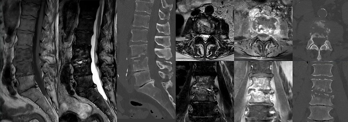 MRI
MRI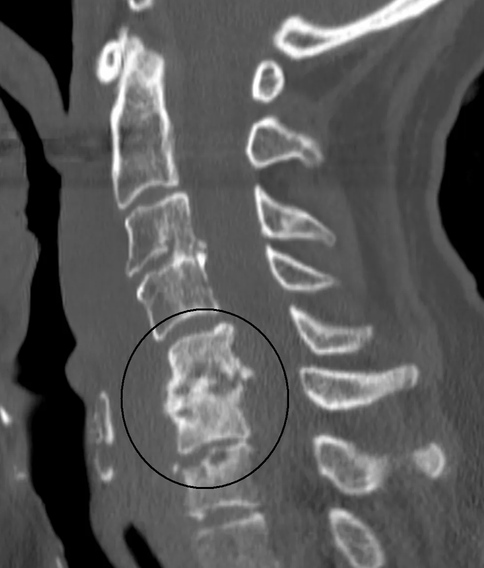 CT
CTDefinition
Spondylodiscitis is an infection of the spine that involves both the vertebral bodies (spondylitis, vertebral osteomyelitis) and the intervertebral disc (discitis). The terms essentially describe stages of the same disease process. It is usually a bacterial (pyogenic) infection; there are also nonpyogenic forms, e.g. due to mycobacteria. Because of its insidious course, the diagnosis is often delayed.
Classification
- Native (spontaneous) spondylodiscitis: without preceding procedure, usually hematogenous.
- Postoperative spondylodiscitis: after spinal procedures, also after lumbar puncture or trauma.
- Pyogenic and nonpyogenic form: the latter mainly due to Mycobacterium tuberculosis, atypical mycobacteria, fungi or Brucella.
- By location: lumbar, thoracic, cervical.
Occurrence & epidemiology
- Native vertebral osteomyelitis accounts for 3–5 % of all cases of osteomyelitis and is the most common form of hematogenous osteomyelitis from age 50 onward.
- Incidence has increased over the past three decades, probably owing to the aging population, more immunocompromised people, more frequent vascular access and better diagnostics.
- Men are affected about 1.5 times as often as women.
- Location: lumbar spine 56.2 %, thoracic spine 18.3 %, cervical spine 6.7 %.
Aetiopathogenesis
- Routes of infection: most commonly hematogenous via the segmental arteries that end in the well-perfused vertebral endplate; typically two adjacent vertebrae are infected and the avascular disc between them secondarily. In children, vessels extend into the disc, which is then the initial site. Less often, direct inoculation (after surgery, lumbar puncture or trauma) or contiguous spread from neighboring structures (e.g. esophageal perforation, diverticular abscess).
- Portals of entry: skin wounds and skin infections, oral cavity, urinary and gastrointestinal tract, endocarditis.
- Pathogens: most commonly Staphylococcus aureus (including MRSA). Coagulase-negative staphylococci and Cutibacterium acnes usually only with implants; in end-stage kidney disease with repeated vascular access also S. epidermidis. Nonpyogenic pathogens (tuberculosis, fungi, Brucella) mainly in immunodeficiency or with corresponding travel and exposure history.
- Risk factors: older age, diabetes mellitus, immunodeficiency including glucocorticoid-induced, advanced liver disease, end-stage kidney disease, alcohol use disorder, obesity, intravenous drug use, indwelling catheters, recent spinal procedures.
Clinical features
- Leading symptom: insidious back pain in about 90 %, often present for several months before medical care is sought; local percussion and pressure tenderness, paravertebral muscle spasm, often continuous.
- Constitutional symptoms such as fever, chills and malaise in about 65 %; however, many patients are afebrile.
- Neurologic deficits in 5–30 %: radicular pain, weakness and sensory disturbances due to compression of the spinal cord or nerve roots; most frequent with thoracic involvement.
- Acute severe pain suggests an epidural abscess or a pathologic vertebral fracture.
- Complications: paravertebral and epidural abscesses (epidural especially cervical), destruction of vertebral bodies with instability and kyphosis, spinal cord syndrome.
- Spinal tuberculosis: typically thoracic or at the thoracolumbar junction with involvement of the vertebral body and often a preserved disc.
Diagnosis
- History and examination: travel, procedures, preceding infections; complete neurologic examination (dermatomes, key muscles, reflexes) to localize the level; search for the source of infection (heart, lungs, abdomen, skin).
- Laboratory tests: ESR, CRP, complete blood count, electrolytes, kidney and liver tests. Leukocytosis is often absent; elevated inflammatory markers are nonspecific. Anemia and thrombocytosis are common.
- Blood cultures: two sets from two different peripheral sites. With risk factors, additionally fungal blood cultures, Brucella serology, interferon-gamma release assay; with drug use, HIV and hepatitis tests.
- Biopsy: if blood cultures are negative, image-guided aspiration or biopsy (sensitivity often below 50 %) for culture and histology (acute or chronic inflammation, granulomas, tumor).
- MRI (most important imaging method): detects the infection earlier than radiographs and CT and shows soft tissues better, including abscesses, muscles and ligaments, spinal cord, cauda equina and nerve roots.
- X-ray: often normal early, disc space narrowing and destruction of the adjacent endplates only after 2–4 weeks; flexion-extension views show deformity and instability.
- CT: precise assessment of bone destruction, pathologic fractures and stability.
- FDG-PET: mainly when MRI is not possible; distinguishes infection from degenerative changes but not reliably from tumors.
Keep learning in the app
Further reading (open access)
Cross-references
Note: Learning content for medical education – not a treatment recommendation and no substitute for diagnosis or treatment decisions in individual cases. Treatment and management are deliberately not covered on this page.