Pneumocystis pneumonia (PCP)
- Synonyms
- PJP, Pneumocystis jirovecii pneumonia, pneumocystosis
- Specialty
- Internal medicine · Infectious diseases
- Images
- CT 1 · Histology 1
- Last updated
- 10/2026 · Dr. Pascal Bafteh
Contents
Images (2)
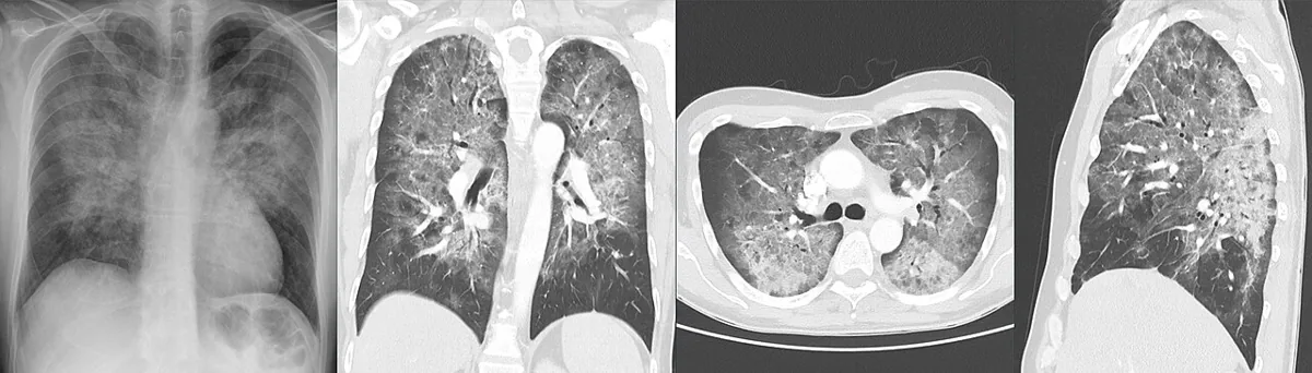 CT
CT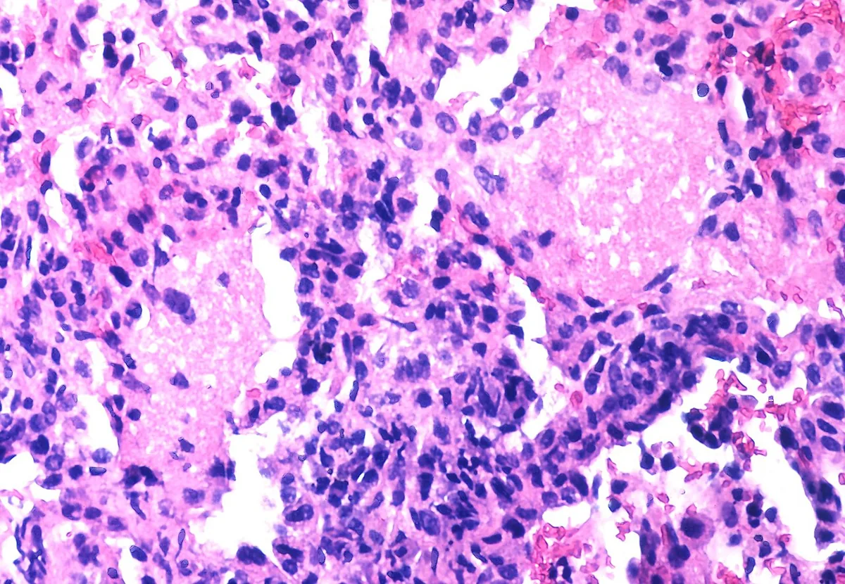 Histology
Definition
Pneumocystis pneumonia (PCP, also PJP) is a pneumonia caused by the atypical fungus Pneumocystis jirovecii (formerly P. carinii). It occurs almost exclusively with impaired cell-mediated immunity and is one of the most important opportunistic infections in advanced HIV infection; in people with HIV it is an AIDS-defining illness.
Occurrence & epidemiology
- P. jirovecii occurs worldwide. Primary infection usually occurs in early childhood; most immunocompetent children have developed specific antibodies by the age of 4 years.
- Since effective HIV medicines became available, the frequency among people with HIV has fallen sharply in well-resourced countries. A high risk persists in undiagnosed HIV infection, especially with CD4 counts below 200/µL or below 14 % (HIV stage 3).
Aetiopathogenesis
- Pathogen: Pneumocystis jirovecii, originally classified as a protozoon, has been considered a fungus since the 1990s based on molecular analyses. Its life cycle consists of trophic forms and cysts.
- Transmission: airborne; causes no disease in immunocompetent people.
- Risk groups: HIV infection with a low CD4 count; organ transplant recipients; hematologic cancers; use of immunosuppressive drugs.
Clinical features
- Leading symptoms: fever, dyspnea (initially on exertion) and dry, nonproductive cough; occasionally chest discomfort.
- Course: insidious over several weeks in HIV infection, faster over a few days in other forms of immunodeficiency.
- Findings: tachypnea and tachycardia, especially on exertion; on auscultation sometimes diffuse fine crackles, but often normal.
- Hypoxemia as the hallmark finding, often with oxygen desaturation on exertion.
- Complications: spontaneous pneumothorax, respiratory failure.
Histology
In tissue, bronchoalveolar lavage or induced sputum, the organizms are found in the alveolar spaces. Giemsa, Diff-Quik and Wright stains show trophic forms and cysts without staining the cyst wall; Grocott-Gomori methenamine silver (GMS), Gram-Weigert, cresyl violet and toluidine blue stains specifically mark the cyst wall. Most stains have low sensitivity; some laboratories additionally use direct immunofluorescence with monoclonal antibodies.
Diagnosis
- Chest X-ray: typically bilateral, diffuse perihilar or fine granular opacities; in 20–30 % the radiograph is normal.
- Chest CT: ground-glass opacities, even with a normal radiograph; a normal CT makes PCP very unlikely.
- Pulse oximetry and arterial blood gas analysis: hypoxemia and increased alveolar-arterial oxygen gradient, sometimes before infiltrates are visible.
- Laboratory tests: elevated serum 1,3-beta-D-glucan and LDH support the suspicion but are nonspecific.
- Pulmonary function testing: reduced diffusing capacity (rarely used for diagnosis).
- Pathogen detection: from induced sputum, bronchoalveolar lavage or tracheal aspirate; bronchoalveolar lavage is considerably more sensitive than induced sputum. Nucleic acid amplification (PCR) is considered the gold standard with the highest yield; in addition immunofluorescence and microscopic stains.
- HIV test in every case of PCP without a known cause of immunodeficiency.
Keep learning in the app
Further reading (open access)
Cross-references
Note: Learning content for medical education – not a treatment recommendation and no substitute for diagnosis or treatment decisions in individual cases. Treatment and management are deliberately not covered on this page.