Cytomegalovirus infection (CMV)
Board exam relevance: in 1 of 105 exam reports · rank 181- Synonyms
- CMV, cytomegaly, human herpesvirus 5, HHV-5
- Specialty
- Internal medicine · Infectious diseases
- Images
- Histology 1 · X-ray 1
- Last updated
- 10/2026 · Dr. Pascal Bafteh
Contents
Images (2)
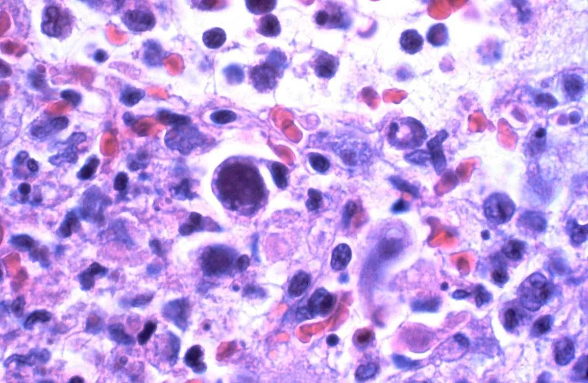 Histology
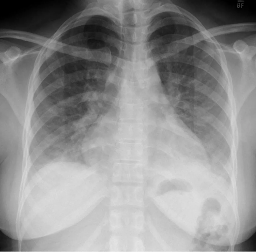 X-ray
X-rayDefinition
Cytomegalovirus infection (CMV infection) is an infection with human herpesvirus 5. Like all herpesviruses, CMV remains latent in the body for life after primary infection and can reactivate. In immunocompetent people the infection is usually unnoticed or presents as a mononucleosis-like illness; in immunodeficiency and after infection in utero it can cause severe organ disease. CMV is the most common viral cause of congenital infection.
Classification
- Primary infection in seronegative people.
- Reactivation of latent virus or reinfection with a different CMV strain.
- CMV infection (virus detection) is distinguished from CMV disease (virus detection with symptoms or organ involvement).
- Congenital, perinatal and postnatal infection.
Occurrence & epidemiology
- Worldwide, about 80 % of adults carry CMV; seroprevalence rises with age and is higher in lower socioeconomic groups and resource-limited regions.
- In Germany, seroprevalence in pregnant women is about 47 %; a study of blood donors found 46 % and of kidney transplant recipients 77 %.
- Young children with congenital or postnatal infection shed large amounts of virus for prolonged periods.
Aetiopathogenesis
- Pathogen: human herpesvirus 5 (betaherpesvirus) with double-stranded DNA; only one serotype but numerous genetically distinct strains. Latency mainly in hematopoietic stem cells and monocytes.
- Transmission: via saliva, urine, tears, genital secretions, breast milk and blood, e.g. through kissing, sexual contact, breastfeeding (in about 35 % of infants of seropositive mothers), blood products and donor organs; also transplacentally and during birth.
- Incubation period: 4–6 weeks in symptomatic primary infection.
- Infectiousness: after reactivation, a seropositive person can shed the virus intermittently for life.
- Risk factors for severe disease: advanced HIV infection, status after transplantation (solid organ or stem cells), other congenital or acquired immunodeficiencies and immunosuppressive drugs.
Clinical features
- Immunocompetent people: usually asymptomatic or nonspecific with fever, fatigue and flu-like symptoms. CMV mononucleosis resembles EBV mononucleosis but usually lacks marked pharyngitis; typical are a mostly subclinical hepatitis without jaundice and atypical lymphocytes.
- After CMV-containing blood products: 2–4 weeks later, fever lasting 2–3 weeks with the picture of CMV mononucleosis.
- Immunodeficiency: usually due to reactivation; pneumonia, hepatitis, gastrointestinal involvement with ulceration of the colon (abdominal pain, bleeding) or esophagus (odynophagia), CNS involvement and retinitis with risk of blindness. In advanced HIV infection, retinitis occurs mainly at very low CD4 counts.
- Pregnancy: about 75 % of primary infections are asymptomatic. Primary infection in the first trimester carries the highest risk for the fetus.
- Congenital infection: growth retardation, hearing loss, microcephaly and late neurologic sequelae; severe liver and CNS damage.
Histology
In tissue infection, enlarged cells with large intranuclear inclusion bodies (owl's eye cells) are seen. Histopathologic evidence is the gold standard for tissue-invasive CMV disease.
Diagnosis
- Serology (immunocompetent people, pregnant women): seroconversion (first appearance of CMV IgG in two samples taken about 2 weeks apart) proves primary infection. IgM alone is not sufficient, as it may also appear with reactivation or other viral infections and can persist for a long time. Low IgG avidity indicates infection within about the past 3 months.
- Distinction from EBV mononucleosis: usually no pharyngitis, negative heterophile antibody test, positive CMV serology.
- Immunosuppressed patients: quantitative detection of CMV DNA (PCR, viral load) or pp65 antigen in leukocytes in blood; high or rising levels suggest invasive disease. Virus detection alone does not prove disease, as it may reflect mere viral shedding.
- Organ involvement: biopsy with histology (e.g. in colitis, esophagitis, pneumonia); ophthalmologic funduscopy if retinitis is suspected.
- Newborns: PCR of saliva or urine within the first days of life; IgM is absent in up to 80 % of congenitally infected infants.
Keep learning in the app
Further reading (open access)
Cross-references
Note: Learning content for medical education – not a treatment recommendation and no substitute for diagnosis or treatment decisions in individual cases. Treatment and management are deliberately not covered on this page.