Benign liver tumors (hemangioma, FNH, adenoma)
Board exam relevance: in 1 of 105 exam reports · rank 181- Synonyms
- liver haemangioma, hepatic hemangioma, FNH, liver adenoma, benign liver lesion, hepatic adenoma
- Specialty
- Internal medicine · Liver & biliary tract
- Images
- Ultrasound 1 · CT 2 · Histology 1
- Last updated
- 10/2026 · Dr. Pascal Bafteh
Contents
Images (4)
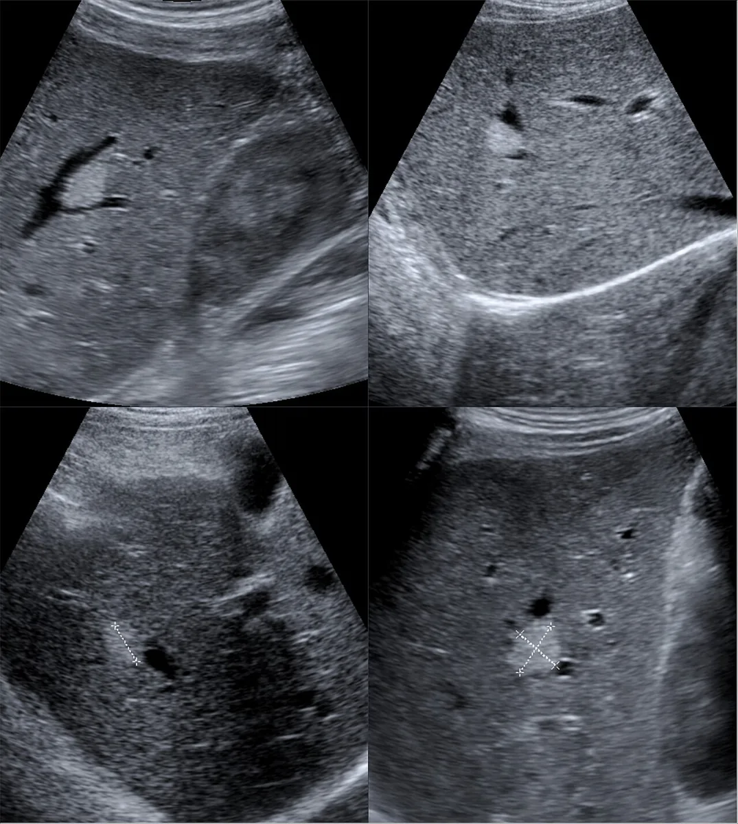 Ultrasound
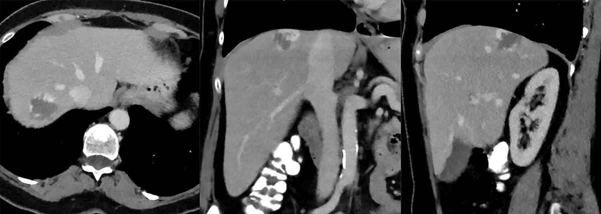 CT
CT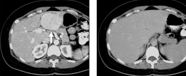 CT
CT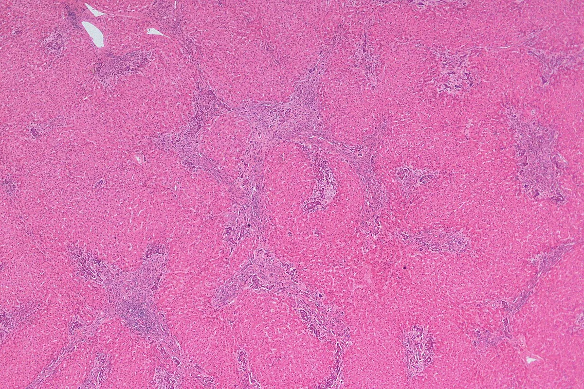 Histology
Definition
Benign liver tumors are non-malignant liver masses. They are relatively common, usually cause no symptoms and are mostly discovered incidentally on ultrasound, CT or MRI. The most important forms are hemangioma, focal nodular hyperplasia (FNH) and hepatocellular adenoma.
Classification
- Hemangioma: benign vascular malformation consisting of blood-filled spaces; the most common benign liver tumor
- Focal nodular hyperplasia (FNH): nodular proliferation of normal liver cells around a central scar with a feeding artery; the second most common solid liver lesion
- Hepatocellular adenoma: benign tumor of liver cells without normal portal tracts; classified into molecular subtypes, including the beta-catenin-activated subtype
- Rarer forms: lipomas, fibrous tumors and bile duct adenomas
Occurrence & epidemiology
Hemangiomas occur in up to 20% of adults, more often in women. Hepatocellular adenomas occur mainly in women of child-bearing age, particularly with use of hormonal contraceptives.
Aetiopathogenesis
Hemangiomas are regarded as congenital vascular malformations. FNH is considered a reactive hyperplasia of liver tissue in response to a local vascular anomaly. Hepatocellular adenomas are associated with estrogens, presumably through their effect on liver cells; further associations exist with androgens and certain metabolic disorders.
Clinical features
- Hemangioma: usually small and asymptomatic; symptoms such as discomfort and fullness are more likely above a size of 4 cm. In infants, hemangiomas often regress spontaneously; however, large hepatic hemangiomas can occasionally cause arteriovenous shunting with heart failure and sometimes consumptive coagulopathy in them.
- FNH: almost always asymptomatic
- Hepatocellular adenoma: usually asymptomatic; large adenomas cause right upper quadrant discomfort. Larger adenomas (typically above 5 cm) can rupture and lead to intraperitoneal hemorrhage with peritonitis and shock. Rarely, adenomas undergo malignant change; in the beta-catenin subtype, the risk of malignant transformation is about 10%.
Liver values are usually normal or only slightly abnormal.
Diagnosis
- Hemangioma: on ultrasound usually a well-defined, hyperechoic lesion; on CT and MRI typically peripheral nodular contrast enhancement with gradual filling from the outside inwards
- FNH: diagnosis usually with MRI or CT with a liver-specific contrast agent; classically a lesion with a central scar
- Hepatocellular adenoma: suspected on ultrasound or CT; MRI with a liver-specific (hepatobiliary) contrast agent is more sensitive than CT, distinguishes adenoma from FNH and can identify subtypes with an increased risk of malignant transformation
- Biopsy: only when imaging does not allow a clear classification
Keep learning in the app
Further reading (open access)
Cross-references
More topics: Liver & biliary tract
Note: Learning content for medical education – not a treatment recommendation and no substitute for diagnosis or treatment decisions in individual cases. Treatment and management are deliberately not covered on this page.