Hepatic encephalopathy
Board exam relevance: in 1 of 105 exam reports · rank 181- Synonyms
- HE, portosystemic encephalopathy, hepatic coma, liver-related confusion, minimal hepatic encephalopathy, flapping tremor
- Specialty
- Internal medicine · Liver & biliary tract
- Images
- MRI 1
- Last updated
- 10/2026 · Dr. Pascal Bafteh
Contents
Images (1)
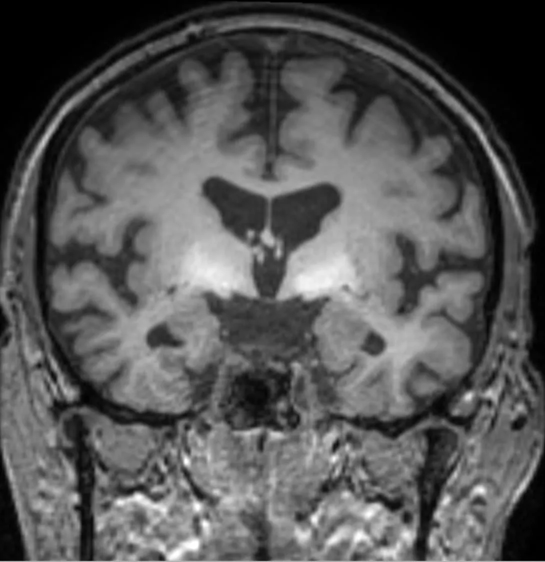 MRI
MRIDefinition
Hepatic encephalopathy (HE) comprises all disorders of central nervous system function that occur as a consequence of acute or chronic liver disease or portosystemic collateral circulation. In liver cirrhosis, the spectrum ranges from deficits detectable only by psychometric testing through increasing psychomotor slowing to coma.
HE is a diagnosis of exclusion, because its symptoms are non-specific.
Classification
HE is graded according to the West Haven criteria; minimal HE is added to these:
- Minimal HE (MHE): clinically normal, deficits only on psychometric or neurophysiological testing
- Grade 1: mental slowing, poor concentration, reduced drive, impaired fine motor skills (e.g. handwriting)
- Grade 2: marked drowsiness, lethargy, disorientation to time, slurred speech, flapping tremor
- Grade 3: somnolence to stupor, disorientation to time and place, incoherent speech, asterixis, rigidity
- Grade 4: coma
Minimal HE and grade 1 are grouped together as covert HE, grades 2–4 as overt HE. By course, episodic HE, recurrent HE (at least two episodes within six months) and persistent HE with continuous symptoms are distinguished.
Occurrence & epidemiology
At the time cirrhosis is diagnosed, 10–21% of patients already have overt HE; over the course of the disease, about 30–45% develop overt HE. Evidence of minimal HE is found in 20–85% of clinically unremarkable patients with cirrhosis.
Aetiopathogenesis
The central cause is the reduced capacity of the cirrhotic liver to detoxify ammonia. In addition, substances produced in the gut reach the circulation unfiltered via portosystemic collaterals. In the brain, ammonia and inflammatory mediators lead to low-grade glial edema with astrocyte dysfunction; changes in GABA neurotransmission also contribute.
Acute episodes are usually provoked by precipitating factors:
- infections (e.g. spontaneous bacterial peritonitis, urinary tract infection, pneumonia)
- gastrointestinal bleeding, for example from varices or ulcers
- electrolyte disturbances, especially hypokalemia and hyponatremia
- fluid depletion (dehydration)
- constipation and very high protein intake
- sedating substances such as alcohol, sedatives or certain analgesics
- acidosis and kidney failure
Clinical features
Early signs are sleep disturbances, poor concentration, slowed thinking, mood changes and deteriorating handwriting. With increasing severity, drowsiness, disorientation, behavioral abnormalities, ataxia and slurred speech follow, up to somnolence and coma.
Asterixis ("flapping tremor") is characteristic: a coarse flapping of the outstretched hands with the wrists dorsiflexed. A sweet, musty breath odour (fetor hepaticus) can occur at any stage. Even minimal HE impairs daily life and quality of life, for example fitness to drive.
Diagnosis
Clinical diagnosis
Overt HE is diagnosed and graded clinically according to the West Haven criteria. Other causes of impaired consciousness are excluded, in particular hypo- or hyperglycemia, electrolyte disturbances, infections of the nervous system, intoxications, Wernicke's encephalopathy and hemorrhage or space-occupying lesions in the brain (cross-sectional imaging).
In every episode, precipitating factors are specifically sought: inflammatory markers and cultures, diagnostic paracentesis, electrolytes and renal function, signs of gastrointestinal bleeding.
Tests and additional investigations
- Minimal HE: detected only by testing, e.g. the Psychometric Hepatic Encephalopathy Score (PHES, a battery of five paper-and-pencil tests) or critical flicker frequency analysis
- Blood ammonia: raised, but correlates poorly with severity and is not diagnostic on its own
- EEG: diffuse slowing even in mild HE, sensitive but non-specific
Keep learning in the app
Further reading (open access)
Cross-references
More topics: Liver & biliary tract
Note: Learning content for medical education – not a treatment recommendation and no substitute for diagnosis or treatment decisions in individual cases. Treatment and management are deliberately not covered on this page.