Primary biliary cholangitis (PBC)
Board exam relevance: in 8 of 105 exam reports · rank 39- Synonyms
- PBC, primary biliary cirrhosis, autoimmune bile duct disease
- Specialty
- Internal medicine · Liver & biliary tract
- Images
- Histology 1 · Blood smear & cytology 1
- Last updated
- 10/2026 · Dr. Pascal Bafteh
Contents
Images (2)
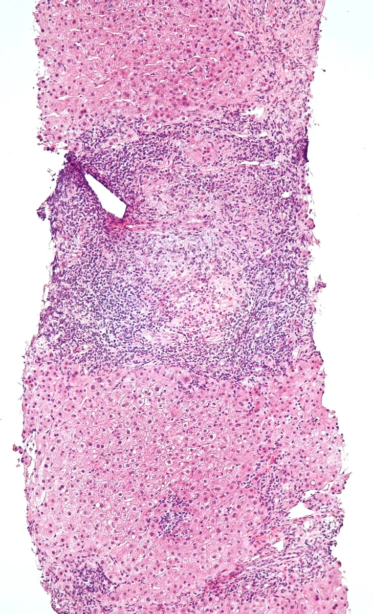 Histology
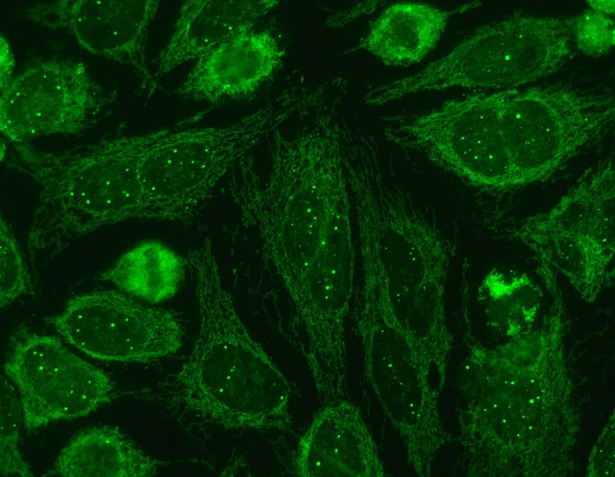 Blood smear & cytology
Definition
Primary biliary cholangitis (PBC) is a chronic, non-suppurative, granulomatous and destructive inflammation of the small intrahepatic bile ducts, presumably of autoimmune origin. It leads to chronic cholestasis and may progress to cirrhosis over years. The former name "primary biliary cirrhosis" was abandoned in 2014 because many patients are now recognized in early stages without cirrhosis; the abbreviation PBC was retained.
Occurrence & epidemiology
About 90–95% of those affected are women, usually aged 40 to 70 years. Prevalence is reported as about 2 to 40 per 100,000 inhabitants; incidence is rising. Men are often less symptomatic.
Aetiopathogenesis
An interplay of genetic factors and environmental influences is assumed; smoking and frequent urinary tract infections are associated with a higher risk of disease. In 90–95%, antimitochondrial antibodies (AMA) against antigens of the inner mitochondrial membrane are found. The immune reaction destroys the epithelium of the interlobular and septal bile ducts, so that bile is retained and the liver is damaged.
Further autoimmune diseases are common, especially Sjögren's syndrome, systemic sclerosis or CREST syndrome and autoimmune thyroiditis.
Clinical features
Many patients are now discovered incidentally because of raised cholestatic enzymes. Up to 80% suffer from fatigue and pruritus, which often occurs before jaundice. Other complaints and findings:
- discomfort in the right upper quadrant, an enlarged firm liver, splenomegaly
- sicca symptoms and arthralgia
- xanthelasmas and hyperpigmentation of the skin
- jaundice as a sign of advanced disease
- fat malabsorption with deficiency of fat-soluble vitamins and osteoporosis
- in late stages, signs of cirrhosis and portal hypertension
Histology
A cholangitis of the interlobular and septal bile ducts is typical. Florid duct lesions with marked periductal inflammation, necrosis and granulomas are considered almost pathognomonic. The infiltrate consists mainly of lymphocytes and mononuclear cells. Over time, bile ducts are lost (ductopenia). Staging follows Ludwig or Scheuer.
Diagnosis
PBC is diagnosed when at least two of three criteria are met:
- chronically raised cholestatic enzymes (especially alkaline phosphatase) for more than 6 months
- antimitochondrial antibodies or PBC-specific antinuclear antibodies
- typical histology
Further findings and investigations:
- ALP and GGT raised, bilirubin usually normal at first; a rise indicates progression
- IgM often raised, cholesterol often raised
- AMA-M2 by ELISA: sensitivity about 90%, specificity about 96%; PBC-specific ANA against sp100 ("nuclear dots") and gp210 (nuclear rim pattern), especially in AMA-negative patients
- ultrasound to exclude biliary obstruction; enlarged lymph nodes at the liver hilum are common
- elastography to estimate the fibrosis stage
- liver biopsy if the diagnosis is unclear or an additional liver disease is suspected
Keep learning in the app
Further reading (open access)
Cross-references
Note: Learning content for medical education – not a treatment recommendation and no substitute for diagnosis or treatment decisions in individual cases. Treatment and management are deliberately not covered on this page.