Liver cirrhosis
Board exam relevance: in 13 of 105 exam reports · rank 17- Synonyms
- cirrhosis, hepatic cirrhosis, end-stage liver disease, scarring of the liver, decompensated cirrhosis
- Specialty
- Internal medicine · Liver & biliary tract
- Images
- Clinical 3 · Gross specimen 2 · CT 1 · Histology 1
- Last updated
- 10/2026 · Dr. Pascal Bafteh
Contents
Images (7)
 Gross specimen
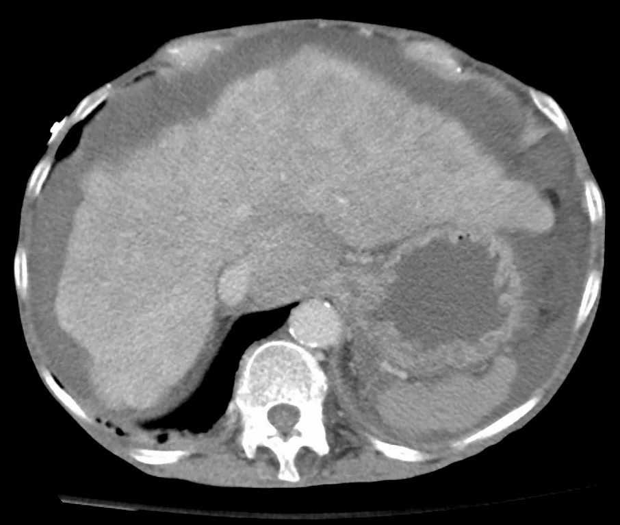 CT
CT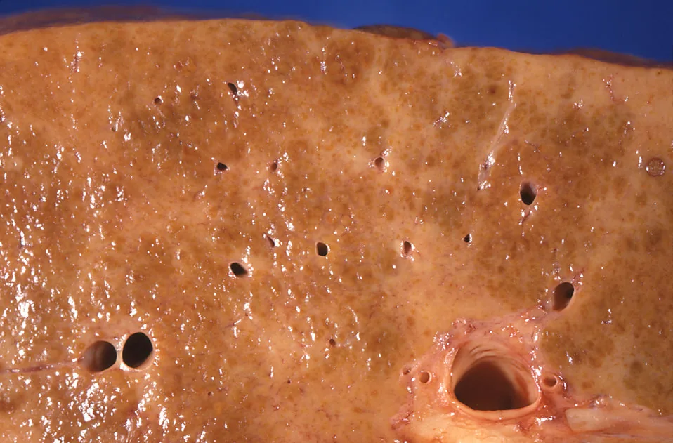 Gross specimen
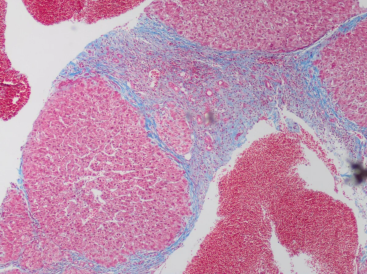 Histology
Definition
Liver cirrhosis is the common end stage of many chronic liver diseases. Regenerative nodules are surrounded by fibrous septa, and the lobular architecture and vascular supply of the liver are lost.
Compensated cirrhosis, which may cause few symptoms for years, is distinguished from decompensated cirrhosis with ascites, variceal bleeding or hepatic encephalopathy.
Classification
The Child-Pugh score assigns 1 to 3 points to each of five criteria:
- Bilirubin: below 2 / 2–3 / above 3 mg/dL
- Albumin: above 3.5 / 2.8–3.5 / below 2.8 g/dL
- INR: below 1.7 / 1.7–2.3 / above 2.3
- Ascites: none / mild / at least moderate
- Hepatic encephalopathy: none / grade 1–2 / grade 3–4
The total defines Child class A (5–6 points), B (7–9) and C (10–15). The MELD score relies only on objective values: MELD-Na uses INR, bilirubin, creatinine and sodium; the current version, MELD 3.0, adds female sex and albumin.
Occurrence & epidemiology
In industrialised countries such as the USA, most cases are due to alcohol, chronic hepatitis C and metabolic dysfunction-associated steatohepatitis (MASH); in parts of Asia and Africa, endemic hepatitis B predominates.
Aetiopathogenesis
- Toxic and metabolic: long-standing excessive alcohol consumption, MASH (formerly NASH)
- Viral: chronic hepatitis B (with or without hepatitis D) and C
- Autoimmune and biliary: autoimmune hepatitis, PBC, PSC, long-standing bile duct obstruction
- Hereditary: hemochromatosis, Wilson's disease, alpha-1 antitrypsin deficiency
- Vascular: Budd-Chiari syndrome, chronic congestive hepatopathy
- Cryptogenic: no cause identifiable
Persistent hepatocellular injury activates the perisinusoidal stellate cells (Ito cells), which produce excess extracellular matrix. Septa and regenerative nodules increase intrahepatic resistance to blood flow, and splanchnic vasodilatation increases inflow. The resulting portal hypertension, together with the loss of liver function, explains most complications.
Clinical features
Symptoms and skin signs
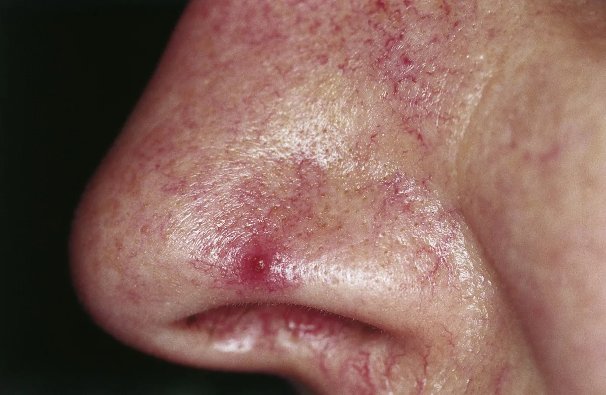
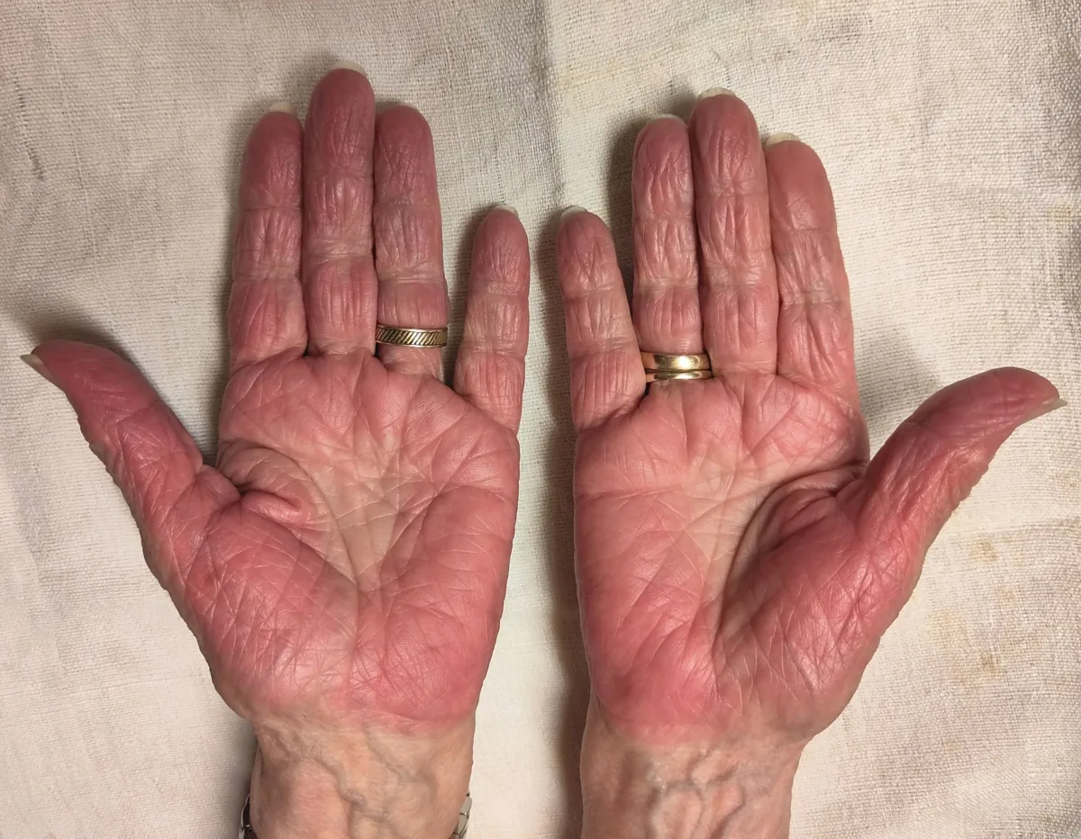
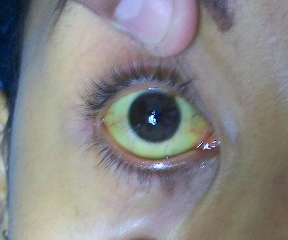
Initially there is often only fatigue, loss of appetite and weight loss. The liver is often palpable as firm with a blunt edge, and in late stages may be small. Signs of chronic liver disease include spider naevi, palmar erythema, white nails, finger clubbing, Dupuytren's contracture, gynecomastia, loss of axillary hair, testicular atrophy and muscle wasting, and in cholestasis jaundice and pruritus.
Complications
- Portal hypertension: esophageal and fundal varices, portal hypertensive gastropathy, splenomegaly with thrombocytopenia, abdominal wall collaterals up to caput medusae
- Ascites, spontaneous bacterial peritonitis and hepatorenal syndrome
- Hepatic encephalopathy ranging from impaired concentration to coma
- Lungs: hepatopulmonary syndrome and portopulmonary hypertension
- Coagulation: reduced clotting factors, yet despite a raised INR a tendency to thrombosis (e.g. portal vein thrombosis)
- Hepatocellular carcinoma: annual incidence depending on the cause about 1% (MASH) up to 3–5% (chronic hepatitis C)
- Acute-on-chronic liver failure (ACLF): acute deterioration with failure of further organs, most often the kidneys
Histology
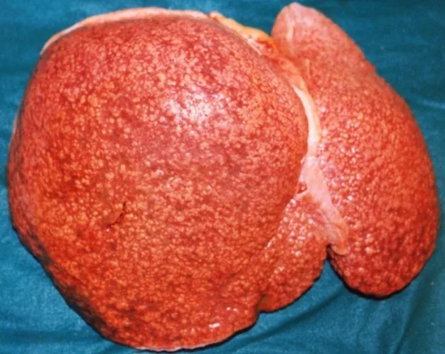
Regenerative nodules and fibrous septa connect portal tracts and central veins. Micronodular cirrhosis shows uniform nodules below 3 mm, macronodular cirrhosis nodules of varying size from 3 mm to 5 cm; mixed forms are common. Fibrosis stages are reported among others according to METAVIR from F0 to F4 (cirrhosis).
Diagnosis
Laboratory tests
- Aminotransferases often only moderately raised or normal; ALP and GGT rise in cholestasis
- Low albumin and raised INR as signs of impaired synthetic function; bilirubin rises as disease progresses
- Thrombocytopenia, leucopenia and anemia
- Pointers to alcohol: AST/ALT ratio above 2, raised GGT, macrocytosis; phosphatidylethanol (PEth) reflects intake over the past 2–4 weeks
- Search for the cause: hepatitis serology, ANA, anti-SMA, IgG, AMA, transferrin saturation, caeruloplasmin, alpha-1 antitrypsin
- Fibrosis scores such as FIB-4 or APRI derived from routine values
Imaging
- Ultrasound: nodular surface, heterogeneous parenchyma, ascites, splenomegaly, collaterals; Doppler for patency and flow of the portal vein
- CT and MRI: nodular liver, varices, portal vein thrombosis, lesions suspicious of HCC with arterial enhancement and washout
- Elastography: transient, shear wave or MR elastography for non-invasive measurement of liver stiffness
Portal hypertension, endoscopy, biopsy
Normal portal pressure is 5–10 mmHg. A hepatic venous pressure gradient (HVPG, measured transjugularly) of 10 mmHg or more defines clinically significant portal hypertension. Non-invasively, a liver stiffness of 25 kPa or more points to it, as do 20–25 kPa with platelets below 150,000/µL or 15–20 kPa with platelets below 110,000/µL. Esophagogastroduodenoscopy shows varices and portal hypertensive gastropathy.
Liver biopsy is regarded as the gold standard but is mainly used when non-invasive findings are unclear or to confirm certain causes; with ascites or coagulopathy, the transjugular route is used.
Keep learning in the app
Further reading (open access)
- MSD Manual Professional: Cirrhosis
- MSD Manual Professional: Hepatic Fibrosis
- MSD Manual Professional: Portal Hypertension
- AWMF-Leitlinienregister 021-017: Komplikationen der Leberzirrhose (S2k-Leitlinie DGVS, Fassung 2019)
- Kim WR et al. 2021: MELD 3.0 – The Model for End-Stage Liver Disease Updated for the Modern Era (Gastroenterology)
- MSD Manual Professional: Ascites
Cross-references
More topics: Liver & biliary tract
Note: Learning content for medical education – not a treatment recommendation and no substitute for diagnosis or treatment decisions in individual cases. Treatment and management are deliberately not covered on this page.