Hemochromatosis
Board exam relevance: in 4 of 105 exam reports · rank 90- Synonyms
- iron overload disease, bronze diabetes, HFE hemochromatosis, iron storage disease
- Specialty
- Internal medicine · Liver & biliary tract
- Images
- Histology 2
- Last updated
- 10/2026 · Dr. Pascal Bafteh
Contents
Images (2)
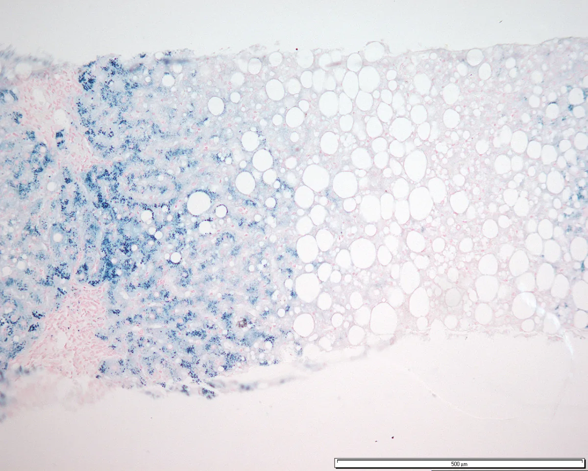 Histology
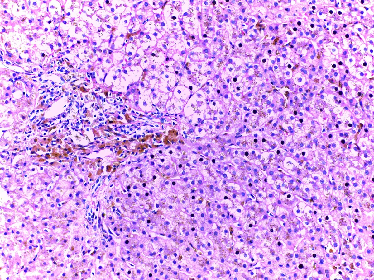 Histology
Definition
Hereditary hemochromatosis is an inherited disorder of iron metabolism with excessive intestinal iron absorption. Iron is deposited in the liver, pancreas, heart, joints, skin and endocrine organs and damages them. Secondary iron overload, for example in people who frequently receive donated blood or with ineffective erythropoiesis, is a separate entity.
Classification
According to the affected gene, four types are distinguished:
- Type 1 (HFE-related): the classic form; more than 80% of cases due to homozygous C282Y mutation; C282Y/H63D compound heterozygosity only rarely leads to relevant iron overload
- Type 2 (juvenile hemochromatosis): mutations in HJV (type 2A) or HAMP (type 2B); onset often in adolescence
- Type 3: mutations in the transferrin receptor 2 gene (TFR2), very rare
- Type 4 (ferroportin disease): autosomal dominant mutations in SLC40A1; in type 4A, transferrin saturation is normal or low
Occurrence & epidemiology
HFE hemochromatosis is inherited in an autosomal recessive manner. Among people of Northern European descent, about 1 in 200 is homozygous and about 1 in 8 heterozygous for the mutation. The mutations are uncommon in people of African descent and rare in people of Asian descent. Penetrance is low: about 70% of C282Y homozygotes have raised ferritin, but only about 10% show organ damage. Men are affected more often and earlier; in women, iron loss through menstruation has a compensating effect until menopause.
Aetiopathogenesis
The central defect is deficient action or production of hepcidin, a peptide hormone produced in the liver. Hepcidin normally inhibits the iron exporter ferroportin and thereby limits iron absorption. Without this brake, the gut absorbs too much iron permanently.
Total body iron is normally about 2.5 g in women and 3.5 g in men; symptoms often appear only when stores exceed 10–20 g. Free iron promotes the formation of reactive oxygen radicals. In the liver, lipid peroxidation and cell death lead to activation of Kupffer and stellate cells, to fibrosis, cirrhosis and an increased risk of hepatocellular carcinoma.
Clinical features
Laboratory abnormalities usually precede symptoms; early symptoms are non-specific. Approximate frequencies in overt disease:
- general symptoms such as weakness and fatigue, and abnormal liver tests: about 75% each
- skin hyperpigmentation: about 70%
- diabetes mellitus ("bronze diabetes"): about 50%
- arthropathy, especially of the second and third metacarpophalangeal joints, often with chondrocalcinosis: about 45%
- erectile dysfunction and hypogonadism: about 45% of men
- cardiomyopathy with heart failure or arrhythmias: about 15%
Liver disease is the most common complication and may progress to cirrhosis; of patients with cirrhosis, 20–30% develop hepatocellular carcinoma. Further findings are hepatomegaly, upper abdominal pain, hypothyroidism and porphyria cutanea tarda.
Histology
In liver biopsy, iron staining (Prussian blue reaction) in HFE hemochromatosis shows iron deposits predominantly in hepatocytes, with a periportal emphasis. In addition, hepatic iron content can be measured quantitatively and the fibrosis stage assessed.
Diagnosis
- Ferritin as the simplest screening test: raised at levels above 200 ng/mL (women) or above 250 ng/mL (men); also raised in inflammation, fatty liver disease, alcohol, tumors or obesity
- Transferrin saturation and fasting serum iron: transferrin saturation usually above 50%, in juvenile hemochromatosis above 90%
- Genetic testing for HFE mutations (C282Y, H63D) confirms type 1 hemochromatosis
- MRI for non-invasive estimation of hepatic iron content, combined with MR elastography for fibrosis assessment
- Liver biopsy with ferritin above 1,000 ng/mL to clarify cirrhosis and in iron overload with a negative genetic test
- Assessment for organ damage: glucose or HbA1c, echocardiography and ECG, hormone status, hand X-rays
Keep learning in the app
Further reading (open access)
Cross-references
More topics: Liver & biliary tract
Note: Learning content for medical education – not a treatment recommendation and no substitute for diagnosis or treatment decisions in individual cases. Treatment and management are deliberately not covered on this page.