Hepatitis A
- Synonyms
- HAV, infectious hepatitis, jaundice, travellers hepatitis, hep A
- Specialty
- Internal medicine · Liver & biliary tract
- Images
- Clinical 1 · Histology 1
- Last updated
- 10/2026 · Dr. Pascal Bafteh
Contents
Images (2)

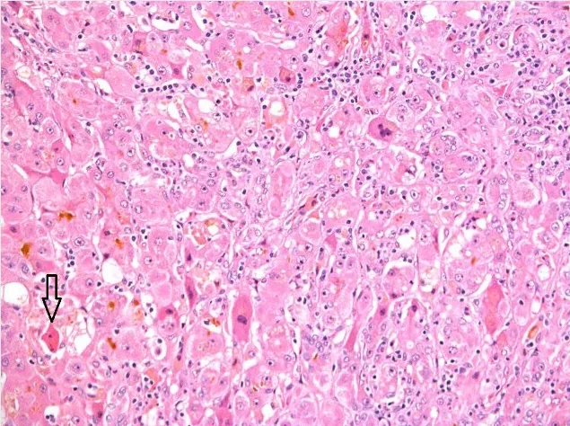 Histology
Definition
Hepatitis A is an acute inflammation of the liver caused by hepatitis A virus (HAV), a single-stranded RNA virus of the family Picornaviridae (genus Hepatovirus). The virus is very stable in the environment and heat-resistant. The disease is exclusively acute; chronic infections do not occur.
Occurrence & epidemiology
HAV occurs worldwide. In countries with intermediate and high endemicity, most people are infected in childhood and adolescence; in some countries more than 90% of children have been exposed to the virus by the age of 10. In industrialised countries the disease has become rare because of high hygiene standards, so many adolescents and adults lack immunity. In Germany, about 30–40% of reported cases before the COVID-19 pandemic were acquired abroad.
Aetiopathogenesis
Humans are the only relevant reservoir. Transmission is fecal-oral:
- as contact or smear infection through close personal contact, for example in households or nurseries, and through sexual contact (especially between men who have sex with men)
- via contaminated drinking water and food, e.g. shellfish, vegetables fertilised with feces or frozen berries
- rarely during the viremic phase via blood or shared drug-use equipment
The incubation period is 15–50 days, on average about 28–30 days. The virus is shed in stool up to about 3 weeks before the onset of jaundice; infectivity is highest in the 2 weeks before jaundice or the rise in aminotransferases. Liver damage results mainly from the immune reaction against infected liver cells.
Clinical features
In young children the infection is usually asymptomatic: below the age of 6, 70–90% of infections are symptom-free, and jaundice is rare. In older children and adults, by contrast, symptomatic acute hepatitis usually develops, with jaundice in more than 70%.
- Prodromal phase: loss of appetite, nausea, vomiting, malaise, occasionally fever
- Icteric phase: yellowing of skin and sclerae, pale stools, dark urine, often pruritus; duration a few days to several weeks
- Findings: enlarged, tender liver with upper abdominal discomfort; splenomegaly in about 25%; occasionally a transient scarlatiniform rash
Most patients recover completely within 2–3 months. About 10–15% develop a protracted cholestatic or biphasic (relapsing) course. Fulminant courses with acute liver failure are rare and mainly affect older people and those with pre-existing liver disease, such as chronic hepatitis B or C. The infection confers lifelong immunity.
Diagnosis
- Laboratory tests: marked rise in aminotransferases, increase in direct and indirect bilirubin and urobilinogen in urine
- IgM anti-HAV: with compatible symptoms, proves recent infection; detectable from the onset of symptoms and positive for about 3–4 months
- IgG anti-HAV (or total anti-HAV): usually already positive at symptom onset; on its own (with negative IgM) a sign of past infection or immunity
- HAV RNA (PCR) in stool or blood: proves recent infection; sequencing of the viral genome is used in specialist laboratories to trace chains of infection in outbreaks
- In severe courses: coagulation (INR) to assess hepatic synthetic function
Keep learning in the app
Further reading (open access)
Cross-references
More topics: Liver & biliary tract
Note: Learning content for medical education – not a treatment recommendation and no substitute for diagnosis or treatment decisions in individual cases. Treatment and management are deliberately not covered on this page.