Budd-Chiari syndrome
Board exam relevance: in 1 of 105 exam reports · rank 181- Synonyms
- hepatic vein thrombosis, hepatic venous outflow obstruction, Budd Chiari, obliterative hepatocavopathy
- Specialty
- Internal medicine · Liver & biliary tract
- Images
- CT 1 · Gross specimen 1
- Last updated
- 10/2026 · Dr. Pascal Bafteh
Contents
Images (2)
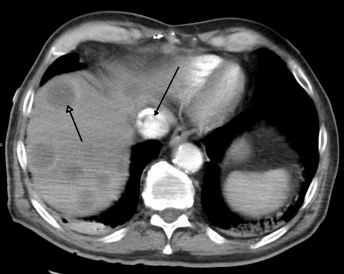 CT
CT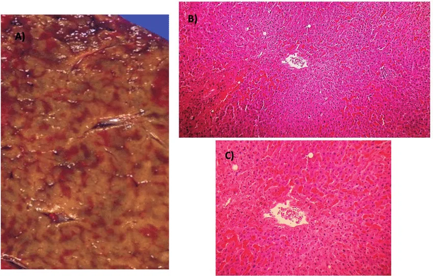 Gross specimen
Definition
Budd-Chiari syndrome is an obstruction of hepatic venous outflow. The obstruction can be located anywhere between the small hepatic veins within the liver and the inferior vena cava up to the right atrium. The congestion damages liver tissue and leads to ascites, hepatomegaly and, over time, to cirrhosis and portal hypertension.
Classification
- Primary: occlusion by thrombosis of the hepatic veins and the adjacent inferior vena cava (most common in Western countries)
- Membranous obstruction: membranes ("webs") in the inferior vena cava above the liver, especially in Asia and South Africa (obliterative hepatocavopathy)
- Secondary: invasion or compression by tumors, cysts or abscesses
By course, acute and chronic forms are distinguished; the syndrome usually develops over weeks to months.
Aetiopathogenesis
Thrombosis usually develops on the basis of an increased tendency to clot:
- Hematological disorders: myeloproliferative neoplasms such as polycythemia vera, as well as paroxysmal nocturnal hemoglobinuria
- Inherited or acquired thrombophilias: protein C, protein S and antithrombin deficiency, factor V Leiden mutation, antiphospholipid syndrome
- Hormonal factors: pregnancy and use of hormonal contraceptives; pregnancy can unmask a previously unnoticed thrombophilia
- Inflammatory diseases: inflammatory bowel disease, Behçet's disease
- Local causes: trauma, infections (e.g. hydatid cyst, amoebiasis), invasion of the hepatic veins by hepatocellular carcinoma or renal cell carcinoma
In some patients the cause remains unclear. Membranous caval obstruction is thought to result from earlier, recanalised thrombosis in adults and from a developmental defect in children.
Clinical features
The spectrum ranges from asymptomatic courses to fulminant liver failure or cirrhosis.
- Acute occlusion: fatigue, right upper quadrant pain, nausea, vomiting, mild jaundice, a tender enlarged liver and ascites; aminotransferases are markedly raised. It typically occurs in pregnancy; fulminant liver failure with encephalopathy is rare.
- Chronic occlusion: often few symptoms for a long time, then fatigue, abdominal pain, hepatomegaly, leg edema and ascites even without cirrhosis; later cirrhosis with variceal bleeding, massive ascites, splenomegaly or hepatopulmonary syndrome.
- Complete occlusion of the inferior vena cava: edema of the abdominal wall and legs and visibly tortuous superficial abdominal veins from the pelvis to the costal margin.
Diagnosis
Budd-Chiari syndrome is suspected with hepatomegaly, ascites, liver failure or cirrhosis without an obvious cause, especially with a known tendency to thrombosis.
- Liver tests: usually abnormal, but with a non-specific pattern
- Doppler ultrasound as the first test: shows the direction of flow and the site of obstruction
- CT and MR angiography if ultrasound is not diagnostic
- Venography with pressure measurement in selected cases
- Liver biopsy occasionally to assess acute stages and cirrhosis
- Search for the cause: thrombophilia testing, blood count and testing for myeloproliferative neoplasms, pregnancy test
Keep learning in the app
Further reading (open access)
Cross-references
More topics: Liver & biliary tract
Note: Learning content for medical education – not a treatment recommendation and no substitute for diagnosis or treatment decisions in individual cases. Treatment and management are deliberately not covered on this page.